. 2015 Mar;81(5):1689-99.
doi: 10.1128/AEM.03610-14. Epub 2014 Dec 29.

Field and experimental evidence of Vibrio parahaemolyticus as the causative agent of acute hepatopancreatic necrosis disease of cultured shrimp (Litopenaeus vannamei) in Northwestern Mexico

Affiliations

Field and experimental evidence of Vibrio parahaemolyticus as the causative agent of acute hepatopancreatic necrosis disease of cultured shrimp (Litopenaeus vannamei) in Northwestern Mexico

Sonia A Soto-Rodriguez et al. Appl Environ Microbiol. 2015 Mar.

Abstract

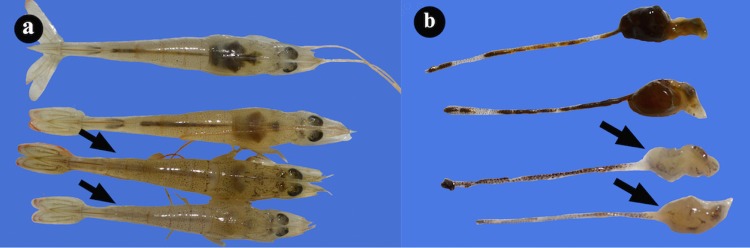
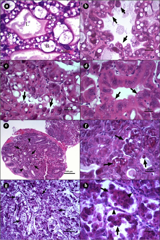
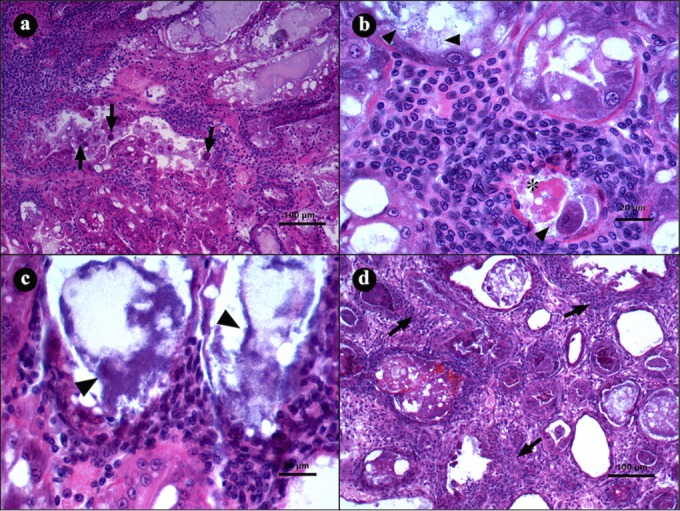
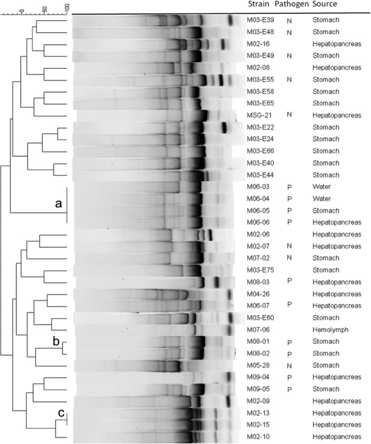
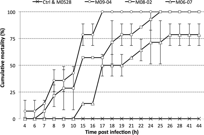
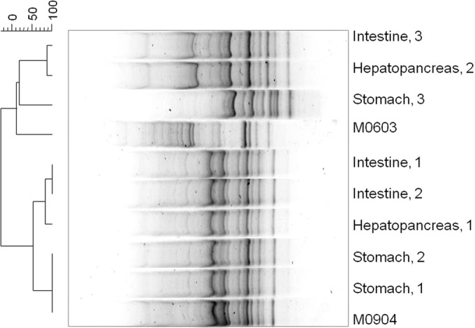
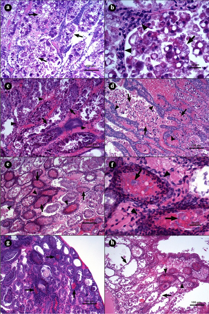
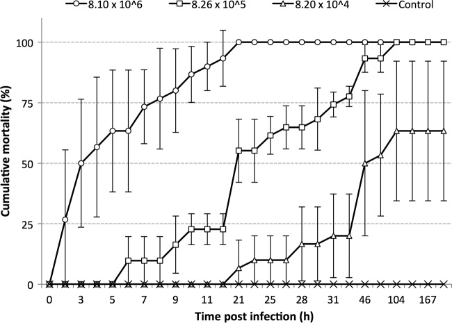
Moribund shrimp affected by acute hepatopancreatic necrosis disease (AHPND) from farms in northwestern Mexico were sampled for bacteriological and histological analysis. Bacterial isolates were molecularly identified as Vibrio parahaemolyticus by the presence of the tlh gene. The tdh-negative, trh-negative, and tlh-positive V. parahaemolyticus strains were further characterized by repetitive extragenic palindromic element-PCR (rep-PCR), and primers AP1, AP2, AP3, and AP and an ems2 IQ2000 detection kit (GeneReach, Taiwan) were used in the diagnostic tests for AHPND. The V. parahaemolyticus strains were used in immersion challenges with shrimp, and farmed and challenged shrimp presented the same clinical and pathological symptoms: lethargy, empty gut, pale and aqueous hepatopancreas, and expanded chromatophores. Using histological analysis and bacterial density count, three stages of AHNPD (initial, acute, and terminal) were identified in the affected shrimp. The pathognomonic lesions indicating severe desquamation of tubular epithelial cells of the hepatopancreas were observed in both challenged and pond-infected shrimp. The results showed that different V. parahaemolyticus strains have different virulences; some of the less virulent strains do not induce 100% mortality, and mortality rates also rise more slowly than they do for the more virulent strains. The virulence of V. parahaemolyticus strains was dose dependent, where the threshold infective density was 10(4) CFU ml(-1); below that density, no mortality was observed. The AP3 primer set had the best sensitivity and specificity. Field and experimental results showed that the V. parahaemolyticus strain that causes AHPND acts as a primary pathogen for shrimp in Mexico compared with the V. parahaemolyticus strains reported to date.


Figures

FIG 1
FIG 1
(a) Photographs of hepatopancreases from healthy L. vannamei shrimp and shrimp naturally infected with AHPND (arrows). (b) The hepatopancreas without external membrane shows atrophy and white color.
FIG 2
FIG 2
Microphotographs of hepatopancreases (HPs) from L. vannamei naturally infected by AHPND. (a) Transversal view of HP with a clear lumen (L), R and B cells, and a normal tubular epithelium. (b to d) Initial stage of infection. (b and c) Transversal view of tubules with elongation of epithelial cells (arrows) toward the lumen. (d) Tubular epithelium with strong reduction of vacuoles in R and B cells (arrows). (e to h) Acute stage of infection. (e) Tubules with a reduction of vacuoles in R and B cells (arrowheads) and severe desquamation of the epithelium (arrows). (f) Magnification of panel e, showing a tubular epithelium necrosis with dead cells inside the lumen (arrows). (g) Severe organ disorganization caused by desquamation of the tubular epithelium. (h) Incipient hemocytic infiltration (arrowhead) in the interstitial spaces of the tubules and loss of continuity of the epithelium due to the necrotic process (arrows). Hematoxylin and eosin staining was used. Microphotographs by S. Abad.
FIG 3
FIG 3
Microphotographs of hepatopancreases (HPs) from L. vannamei naturally infected by AHPND in the terminal stage of infection. (a) Transversal view of HP with severe hemocytic infiltration in the interstitial spaces as a response to epithelium necrosis (arrows). (b and c) Hepatopancreas tubule shows hemocytic infiltration, with necrotic cells (asterisk) and bacterial masses inside the lumen (arrowheads). (d) Transverse view of HP with severe hemocytic infiltration (arrow) and melanization of necrotic material inside the tubules. Hematoxylin and eosin staining was used. Microphotographs by S. Abad.
FIG 4
FIG 4
ERIC-PCR fingerprinting of Vibrio parahaemolyticus isolated from shrimp and water from ponds affected by AHPND. N, nonpathogenic; P, pathogenic. Letters in the dendrogram denote clonal groups (>95%). Band position tolerance, 1%; optimization, 0.2%.
FIG 5
FIG 5
Example of cumulative mortality of juvenile shrimp challenged with Vibrio parahaemolyticus strains. M05-28, 1.20 × 10⁷ CFU ml−1; M09-04, 2.20 × 10⁶ CFU ml−1; M08-02, 3.30 × 10⁶ CFU ml−1; M06-07, 7.82 × 10⁶ CFU ml−1. Control, TSB plus 2.0% NaCl. Bars indicate standard deviations.
FIG 6
FIG 6
DNA fingerprinting of bacterial isolates obtained from challenged shrimp infected with the pathogenic V. parahaemolyticus strain M09-04. The organ of isolation and number of shrimp samples are shown. M09-04, strain inoculated; M06-03, strains not inoculated but used as outgroup in the GTG5 PCR. Band position tolerance, 0.5%; optimization, 0.13%.
FIG 7
FIG 7
Microphotographs of L. vannamei hepatopancreases (HPs) in the acute (a to d) and terminal (e to h) stage of AHPND. (a and b) Hepatopancreas from a shrimp challenged with strain M09-05 at 3 h p.i., showing a general disorganization of the tubules due the epithelium necrosis; the lumen is full of dead cells produced by the epithelium desquamation. (b) Magnification of panel a. Epithelium desquamation and hemocytic infiltration were observed (arrowhead). (c) Hepatopancreas from a shrimp challenged with strain M08-01 at 10 h p.i., showing hemocytic infiltration (arrowhead) and reduction of R and B vacuole cells (arrow). (d) At 16 h p.i. with the same strain, the HP had a larger hemocytic infiltration (arrowhead) and accumulations of necrotic cells inside the tubular lumen (arrow). (e and f) Hepatopancreas from a shrimp challenged with strain M06-06 at 24 h p.i., with severe hemocytic infiltration (arrowhead), epithelium necrosis, deposits of melanin inside the tubular lumen, and bacterial masses (arrow). (g) Hepatopancreas from a shrimp challenged with strain M09-05 at 28 h p.i., with the largest melanization of necrotic tissue inside the tubular lumen (arrow). (h) Hepatopancreas from a shrimp challenged with strain M06-05 at 41 h p.i., showing necrotic tubules without epithelium (arrow) and tubules with hemocytic capsules (arrowhead). Hematoxylin and eosin staining was used. Microphotographs by S. Abad.
FIG 8
FIG 8
Cumulative mortality of juvenile shrimp challenged with Vibrio parahaemolyticus strain M09-04 at different bacterial densities (CFU ml−1). Bars indicate standard deviations.

Similar articles

Cited by

References

    1. Lightner DV. 1999. The Penaeid Shrimp Viruses TSV, IHHNV, WSSV, and YHV: current status in the Americas, available diagnostic methods, and management strategies. J Appl Aquacult 9:27–52. doi:10.1300/J028v09n02_03. - DOI
    1. Del Rio-Rodriguez RE, Soto-Rodriguez S, Lara-Flores M, Cu-Escamilla AD, Gomez-Solano MI. 2006. A necrotizing hepatopancreatitis (NHP) outbreak in a shrimp farm in Campeche, Mexico: a first case report. Aquaculture 255:606–209. doi:10.1016/j.aquaculture.2005.12.014. - DOI
    1. Nunan L, Pantoja C, Gomez-Jimenez S, Lightner D. 2013. “Candidatus hepatobacter penaei,” an intracellular pathogenic enteric bacterium in the hepatopancreas of the marine shrimp Penaeus vannamei (Crustacea: Decapoda). Appl Environ Microbiol 79:1407–1409. doi:10.1128/AEM.02425-12. - DOI - PMC - PubMed
    1. Soto-Rodriguez SA, Gomez Gil B, Roque A., Lozano R. 2010. Density of vibrios in hemolymph and hepatopancreas of diseased Pacific white shrimp, Litopenaeus vannamei, from northwestern Mexico. J World Aquacult Soc 41:76–83. doi:10.1111/j.1749-7345.2009.00335.x. - DOI
    1. Jiravanichpaisal P, Miyazaki Limsuwan TC. 1994. Histopathology, biochemistry and pathogenicity of Vibrio harveyi infecting black tiger prawn, Penaeus monodon. J Aquat Anim Health 6:27–35. doi:10.1577/1548-8667(1994)006<0027:HBAPOV>2.3.CO;2. - DOI

Publication types

MeSH terms