. 2015 Feb 13;290(7):4422-31.
doi: 10.1074/jbc.M114.597518. Epub 2014 Dec 29.

Induction and activation of latent transforming growth factor-β1 are carried out by two distinct domains of pregnancy-specific glycoprotein 1 (PSG1)

Affiliations

Induction and activation of latent transforming growth factor-β1 are carried out by two distinct domains of pregnancy-specific glycoprotein 1 (PSG1)

Angela Ballesteros et al. J Biol Chem. .

Abstract

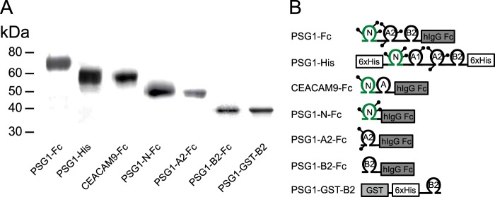
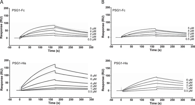
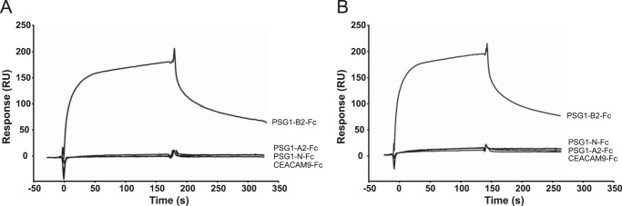
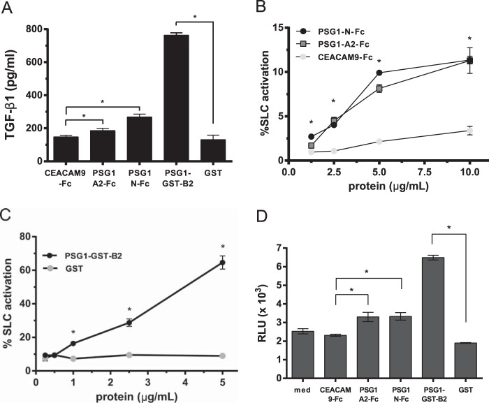
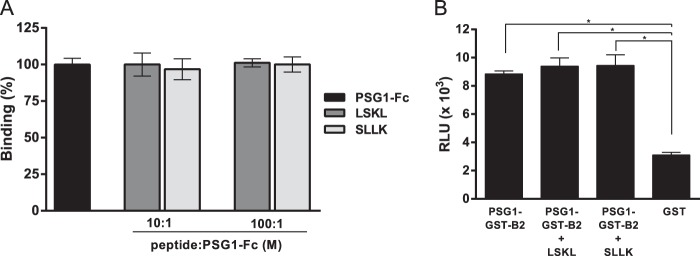
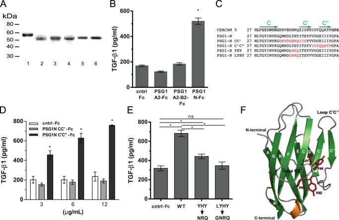
Pregnancy-specific glycoproteins (PSGs) are a family of Ig-like proteins secreted by specialized placental cells. The PSG1 structure is composed of a single Ig variable region-like N-terminal domain and three Ig constant region-like domains termed A1, A2, and B2. Members of the human and murine PSG family have been shown to induce anti-inflammatory cytokines from monocytes and macrophages and to stimulate angiogenesis. We recently showed that recombinant forms of PSG1 (PSG1-Fc and PSG1-His) and PSG1 purified from the serum of pregnant women are associated with the immunoregulatory cytokine TGF-β1 and activated latent TGF-β1. Here, we sought to examine the requirement of specific PSG1 domains in the activation of latent TGF-β1. Plasmon surface resonance studies showed that PSG1 directly bound to the small latent complex and to the latency-associated peptide of TGF-β1 and that this binding was mediated through the B2 domain. Furthermore, the B2 domain alone was sufficient for activating the small latent complex. In separate experiments, we found that the PSG1-mediated induction of TGF-β1 secretion in macrophages was dependent on the N-terminal domain. Mutagenesis analysis revealed that four amino acids (LYHY) of the CC' loop of the N-terminal domain were required for induction of latent TGF-β1 secretion. Together, our results show that two distinct domains of PSG1 are involved in the regulation of TGF-β1 and provide a mechanistic framework for how PSGs modulate the immunoregulatory environment at the maternal-fetal interface for successful pregnancy outcome.

Keywords: Heparan Sulfate; Latency-associated Peptide; Latent TGF-β1; Macrophage; Placenta; Pregnancy; Surface Plasmon Resonance (SPR).


Figures

FIGURE 1.
FIGURE 1.
Schematic of recombinant PSG1 and PSG1 domain constructs. A, PSG1-Fc, PSG1-His, CEACAM9-Fc, PSG1-N-Fc, PSG1-A2-Fc, and PSG1-B2-Fc produced in CHO-K1 cells and PSG1-GST-B2 expressed in bacteria were purified and loaded onto a 4–20% NuPAGE gel. The gel was stained with GelCode Blue to visualize the proteins. B, schematic representation of the proteins listed in A. The position of protein tags (Fc or His) is indicated; lollipops represent potential N-linked glycosylation sites.
FIGURE 2.
FIGURE 2.
PSG1 interacts with SLC and LAP of TGF-β1. Shown are the results from SPR analysis of the interaction of PSG1-Fc and PSG1-His with SLC (A) or LAP (B) of TGF-β1. A protein concentration that ranged from 8 to 0.5 μm was analyzed across blank, SLC, and LAP biosensor surfaces. The data were analyzed using a simultaneous fit algorithm to calculate the kinetic parameters presented in Table 1. Representative SPR sensorgrams for each response are shown as gray lines, and fit analyses are shown as black lines. RU, response units.
FIGURE 3.
FIGURE 3.
The B2 domain of PSG1 interacts with SLC and LAP of TGF-β1. Show are Biacore sensorgrams for the interaction of PSG1-Fc domains and CEACAM9-Fc at 2 μm with SLC (A) and LAP (B) of TGF-β1. Purified CEACAM9-Fc and PSG1-Fc proteins containing the N-terminal, A2, and B2 domains of PSG1 were injected over a CM5 sensor chip with immobilized SLC or LAP of TGF-β1. A flow cell to which no protein was coupled served as a surface control. Representative sensorgrams are shown. RU, response units.
FIGURE 4.
FIGURE 4.
The B2 domain of PSG1 is sufficient for activation of SLC of TGF-β1. A, 2.5 μg/ml CEACAM9-Fc, PSG1-A2-Fc, PSG1-N-Fc, PSG1-GST-B2, or GST was incubated with 50 ng/ml SLC for 1 h at 37 °C and then analyzed for active TGF-β1 by ELISA as described under “Experimental Procedures.” B, increasing concentrations of PSG1-N-Fc, PSG1-A2-Fc, and CEACAM9-Fc were incubated with 50 ng/ml SLC for 1 h at 37 °C, and the percentage of SLC activated was measured with a TGF-β receptor II-Fc capture ELISA. 100% SLC activation was defined as the amount of active TGF-β1 measured following acid activation of 50 ng/ml SLC. C, increasing concentrations of PSG1-GST-B2 or GST were incubated with 50 ng/ml SLC for 1 h at 37 °C, and the percent SLC activated was measured as described for B. D, 2.5 μg/ml CEACAM9-Fc, PSG1-A2-Fc, PSG1-N-Fc, PSG1-GST-B2, GST, or medium alone (med) was incubated with 50 ng/ml SLC in DMEM and 0.1% insulin/transferrin/sodium selenite medium for 1 h at 37 °C and then added in triplicate to luciferase reporter cells (MLECs). The treated MLECs were incubated for 16 h at 37 °C and lysed, and the luciferase activity was analyzed on a GloMax luminometer. The relative light units (RLU) reflect the dose-dependent increase in luciferase activity following active TGF-β signaling. For A–D, all treatments were performed in triplicate for a minimum of three independent experiments. *, p ≤ 0.02 by Student's t test for data in A–D.
FIGURE 5.
FIGURE 5.
The LSKL peptide does not inhibit PSG1-Fc binding to LAP or the activation of SLC via PSG1-GST-B2. A, PSG1-Fc protein (2 μm) was incubated with the LSKL peptide or a control peptide (SLLK) at a 10:1 or 100:1 peptide/protein molar ratio at 37 °C for 1 h and injected at 10 μl/min over a LAP biosensor surface. The percentage of binding of PSG1-Fc in the presence of peptide is shown relative to PSG1-Fc binding to LAP in the absence of peptide (100%). The mean ± S.D. for three independent experiments is shown. B, SLC (50 ng/ml) was incubated with PSG1-GST-B2 alone or with PSG1-GST-B2 in the presence of a 10-fold molar excess of the LSKL or SLLK peptide at 37 °C for 1 h and added in triplicate to luciferase reporter cells (MLECs). After 16 h at 37 °C, the cells were lysed, and the luciferase activity was analyzed on a luminometer. The relative light units (RLU) reflect the increase in luciferase activity following active TGF-β signaling. *, p < 0.002 with Student's t test.
FIGURE 6.
FIGURE 6.
Amino acids in the CC′ loop region of the N-terminal domain of PSG1 are essential for induction of TGF-β1 from RAW264.7 macrophages. A, recombinant proteins used for these studies were separated on a 4–20% NuPAGE gel and stained with GelCode Blue. Lane 1, PSG1-A2-B2-Fc; lane 2, PSG1-N-Fc; lane 3, PSG1-N-CC′-Fc; lane 4, PSG1-N-C′C″-Fc; lane 5, PSG1-N-YHY-Fc; lane 6, PSG1-N-LYHY-Fc. B, TGF-β1 secretion following incubation of RAW264.7 murine macrophages with 3 μg/ml control Fc protein (cntrl-Fc), PSG1-A2-Fc, and PSG1-N-Fc or 6 μg/ml PSG1-A2-B2-Fc to achieve concentrations equimolar to the single-domain proteins. C, sequence alignment of the CC′ and C′C″ loop regions of CEACAM5, PSG1, and PSG1 N-terminal domain mutants. Residues conserved in CEACAM5 and PSG1 are highlighted in gray. The C, C′, and C″ β-strands are indicated with green arrows above the corresponding sequence. Residues in the PSG1 N-terminal domain mutated to the residues corresponding to the same position in CEACAM5 are colored red. D, TGF-β1 secretion of RAW264.7 macrophages following incubation with increasing concentrations of PSG1-N-CC′-Fc, PSG1-N-C′C″-Fc, or control Fc protein. E, RAW264.7 macrophages were incubated with 12 μg/ml wild-type PSG1 N-terminal domain (WT), PSG1-N-Y42N/H43R/Y44Q mutant (YHYNRQ), PSG1-N-L41G/Y42N/H43R/Y44Q mutant (LYHYGNRQ), or control Fc protein. For B–E, TGF-β1 following acid activation was measured in the supernatants by ELISA as described under “Experimental Procedures.” *, p ≤ 0.05 by Student's t test; ns, not significant. F, ribbon representation of the structural model of the PSG1 N-terminal domain. β-Sheets are shown in green and labeled with uppercase white letters. The α-helix between strands E and F is shown in orange, and loops are shown in gray. Residues 41LYHY44 of the CC′ loop are labeled and highlighted in red, and their side chains are shown in stick representation. The CC′ and C′C″ loops and the N and C termini of the protein domain are indicated.

References

    1. Teglund S., Olsen A., Khan W. N., Frängsmyr L., Hammarström S. (1994) The pregnancy-specific glycoprotein (PSG) gene cluster on human chromosome 19: fine structure of the 11 PSG genes and identification of 6 new genes forming a third subgroup within the carcinoembryonic antigen (CEA) family. Genomics 23, 669–684 - PubMed
    1. McLellan A. S., Fischer B., Dveksler G., Hori T., Wynne F., Ball M., Okumura K., Moore T., Zimmermann W. (2005) Structure and evolution of the mouse pregnancy-specific glycoprotein (Psg) gene locus. BMC Genomics 6, 4. - PMC - PubMed
    1. Shanley D. K., Kiely P. A., Golla K., Allen S., Martin K., O'Riordan R. T., Ball M., Aplin J. D., Singer B. B., Caplice N., Moran N., Moore T. (2013) Pregnancy-specific glycoproteins bind integrin αIIbβ3 and inhibit the platelet-fibrinogen interaction. PLoS ONE 8, e57491. - PMC - PubMed
    1. Snyder S. K., Wessner D. H., Wessells J. L., Waterhouse R. M., Wahl L. M., Zimmermann W., Dveksler G. S. (2001) Pregnancy-specific glycoproteins function as immunomodulators by inducing secretion of IL-10, IL-6 and TGF-β1 by human monocytes. Am. J. Reprod. Immunol. 45, 205–216 - PubMed
    1. Martínez F. F., Knubel C. P., Sánchez M. C., Cervi L., Motrán C. C. (2012) Pregnancy-specific glycoprotein 1a activates dendritic cells to provide signals for Th17-, Th2-, and Treg-cell polarization. Eur. J. Immunol. 42, 1573–1584 - PubMed

Publication types

MeSH terms

LinkOut - more resources