. 2014 Dec 21;20(47):17894-904.
doi: 10.3748/wjg.v20.i47.17894.

HIF-1α induces VE-cadherin expression and modulates vasculogenic mimicry in esophageal carcinoma cells

Affiliations

HIF-1α induces VE-cadherin expression and modulates vasculogenic mimicry in esophageal carcinoma cells

Na-Na Tang et al. World J Gastroenterol. .

Abstract

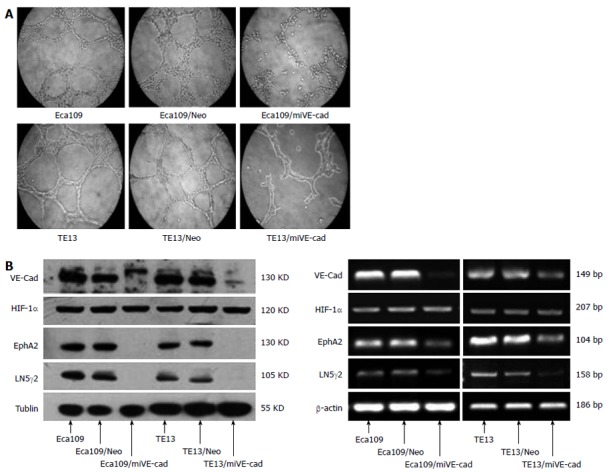
Aim: To investigate whether hypoxia inducible factor (HIF)-1α modulates vasculogenic mimicry (VM) by upregulating VE-cadherin expression in esophageal squamous cell carcinoma (ESCC).

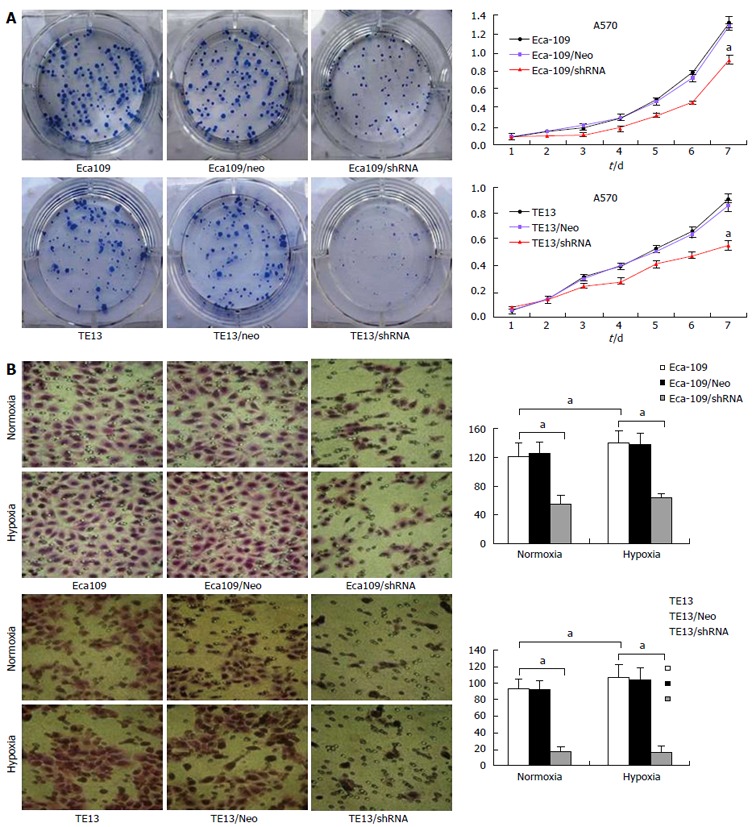
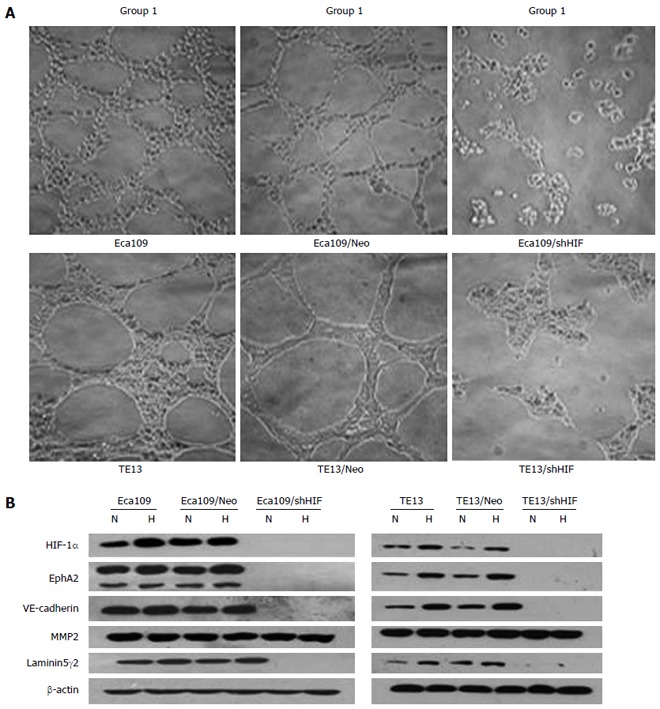
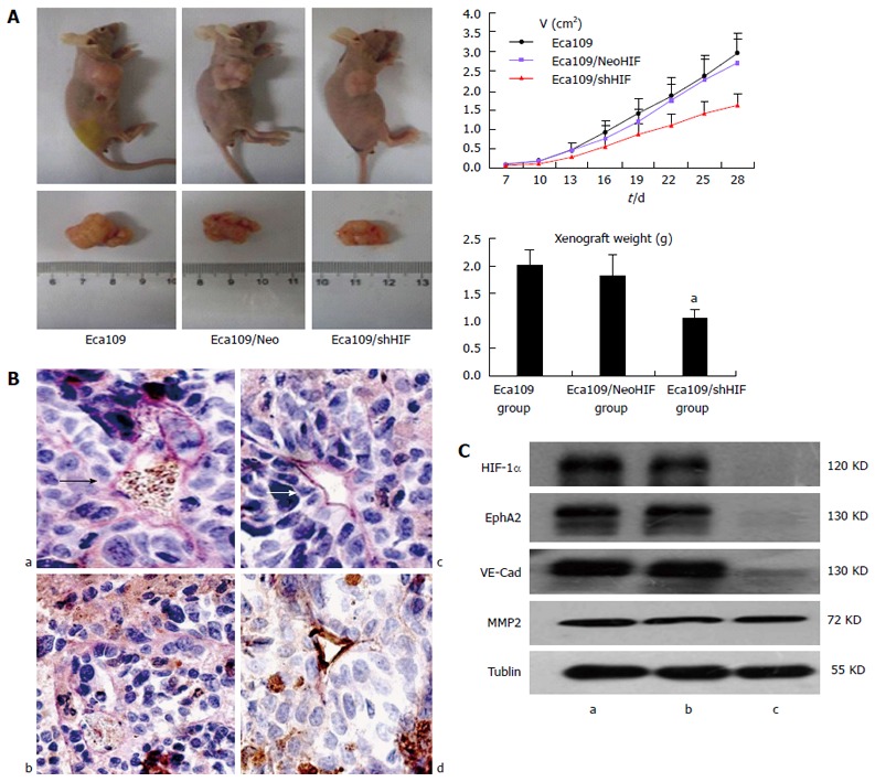
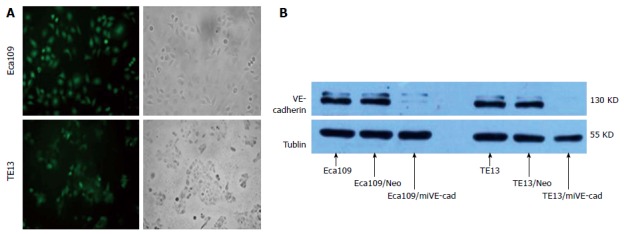
Methods: Esophageal squamous cancer cell lines Eca109 and TE13 were transfected with plasmids harboring small interfering RNAs targeting HIF-1α or VE-cadherin. The proliferation and invasion of esophageal carcinoma cells were detected by MTT and Transwell migration assays. The formation of tubular networks of cells was analyzed by 3D culture in vitro. BALB/c nude mice were used to observe xenograft tumor formation. The relationship between the expression of HIF-1α and VE-cadherin, ephrinA2 (EphA2) and laminin5γ2 (LN5γ2) was measured by Western blot and real-time polymerase chain reaction.

Results: Knockdown of HIF-1α inhibited cell proliferation (32.3% ± 6.1% for Eca109 cells and 38.6% ± 6.8% for TE13 cells, P < 0.05). Both Eca109 and TE13 cells formed typical tubular networks. The number of tubular networks markedly decreased when HIF-1α or VE-cadherin was knocked down. Expression of VE-cadherin, EphA2 and LN5γ2 was dramatically inhibited, but the expression of matrix metalloproteinase 2 had no obvious change in HIF-1α-silenced cells. Knockdown of VE-cadherin significantly decreased expression of both EphA2 and LN5γ2 (P < 0.05), while HIF-1α expression was unchanged. The time for xenograft tumor formation was 6 ± 1.2 d for Eca109 cells and Eca109 cells transfected with HIF-1α Neo control short hairpin RNA (shRNA) vector, and 8.4 ± 2.1 d for Eca109 cells transfected with an shRNA against HIF-1α. Knockdown of HIF-1α inhibited vasculogenic mimicry (VM) and tumorigenicity in vivo.

Conclusion: HIF-1α may modulate VM in ESCC by regulating VE-cadherin expression, which affects VM formation through EphA2 and LN5γ2.

Keywords: Esophageal squamous cell carcinoma; Hypoxia-inducible factor-1α; RNA interference; VE-cadherin; Vasculogenic mimicry.


Figures

Figure 1
Figure 1
Effects of hypoxia inducible factor-1α knockdown on regulation of esophageal cancer cell viability and migration. A: MTT assay demonstrated that Eca109/HIF-1α shRNA and TE13/HIF-1α shRNA cells proliferated slower than normal Eca109 and TE13 cells or Eca109 HIF-1α Neo and TE13 HIF-1α Neo cells; B: Transwell migration assay demonstrated that Eca109/HIF-1α shRNA and TE13/HIF-1α shRNA cells invaded slower than normal Eca109 and TE13 cells or Eca109 HIF-1α Neo and TE13 HIF-1α Neo cells. Expression of HIF-1α and the four VM-related genes in TE13/shHIF and Eca109/shHIF cells under hypoxia vs under normoxia, aP < 0.05. HIF: Hypoxia inducible factor.
Figure 2
Figure 2
Knockdown of hypoxia inducible factor-1α inhibits vasculogenic mimicry formation and expression of vasculogenic mimicry-related genes in vitro. A: Effect of silencing HIF-1α on the formation of VM in ESCC cells (magnification × 200) (Group 1: Untransfected cells; Group 2: Cells transfected with empty vector; Group 3: Cells transfected with pGCsi-HIF3); Control group vs parental cells, P < 0.05; B: Expression of HIF-1α protein and VM-related genes in esophageal squamous cancer cells. Expression of HIF-1α and the four VM-related genes in TE13/shHIF and Eca109/shHIF cells under hypoxia vs under normoxia, P < 0.05. HPF: High-power field; HIF: Hypoxia inducible factor; VM: Vasculogenic mimicry; ESCC: Esophageal squamous cell carcinoma. N: Normoxia; H: Hypoxia.
Figure 3
Figure 3
In vivo effects of hypoxia inducible factor-1α knockdown on regulation of esophageal cancer cell xenograft formation and gene expression. A: Tumor size curve and weight of xenograft (Eca109/shHIF-1α cells vs Eca109 cells and Eca109/HIF-1α Neo cells, aP < 0.05); B: VM structure (arrow) in xenografts of three groups (a: Eca109 group; b: Eca109/shHIF group; c: Eca109/NeoHIF group; d: Normal vessels; arrow, VM structure); C: Western blot detected HIF-1α and EphA2, VE-cadherin and MMP2 expression (a: Eca109 group; b: Eca109/NeoHIF group; c: Eca109/shHIF group; Eca109/shHIF-1α cells vs Eca109 and Eca109 HIF-1α Neo control cells, P < 0.05). HIF: Hypoxia inducible factor; VM: Vasculogenic mimicry.
Figure 4
Figure 4
Identification of esophageal squamous cell carcinoma cells stably transfected with pGCsi-VE-cadherin. A: Expression of green fluorescent protein in two stably transfected ESCC cell lines (magnification × 200); B: Expression of VE-cadherin protein in the two ESCC cell lines. ESCC: Esophageal squamous cell carcinoma.
Figure 5
Figure 5
Knockdown of VE-cadherin inhibits proliferation and promotes apoptosis of esophageal squamous cell carcinoma cells. A: shRNA-mediated VE-cadherin knockdown inhibits the proliferation of ESCC cells. Eca109, TE13 and their derived stable cells were cultured under normoxic conditions and proliferation was determined by MTT assay; aP < 0.05; B: shRNA-mediated VE-cadherin knockdown promoted apoptosis of ESCC cells. Eca109, TE13 and their derived stable cells were cultured under normoxic or hypoxic conditions and apoptosis was detected by Annexin V staining. ESCC: Esophageal squamous cell carcinoma.
Figure 6
Figure 6
shRNA against VE-cadherin inhibits vasculogenic mimicry formation and expression of vasculogenic mimicry-related genes in vitro. A: Effect of silencing VE-cadherin on the formation of VM in ESCCs (magnification × 400); B: Protein and mRNA expression of VE-cadherin and VM-related genes in esophageal squamous cancer cells. ESCC: Esophageal squamous cell carcinoma; VM: Vasculogenic mimicry; VE-cad: VE-cadherin; LN5γ2: Laminin 5γ2; HIF: Hypoxia inducible factor; EphA2: EphrinA2.

References

    1. Zhu P, Ning Y, Yao L, Chen M, Xu C. The proliferation, apoptosis, invasion of endothelial-like epithelial ovarian cancer cells induced by hypoxia. J Exp Clin Cancer Res. 2010;29:124. - PMC - PubMed
    1. Michaylira CZ, Nakagawa H. Hypoxic microenvironment as a cradle for melanoma development and progression. Cancer Biol Ther. 2006;5:476–479. - PubMed
    1. Maniotis AJ, Folberg R, Hess A, Seftor EA, Gardner LM, Pe’er J, Trent JM, Meltzer PS, Hendrix MJ. Vascular channel formation by human melanoma cells in vivo and in vitro: vasculogenic mimicry. Am J Pathol. 1999;155:739–752. - PMC - PubMed
    1. Grunewald TG, Herbst SM, Heinze J, Burdach S. Understanding tumor heterogeneity as functional compartments--superorganisms revisited. J Transl Med. 2011;9:79. - PMC - PubMed
    1. Sun W, Shen ZY, Zhang H, Fan YZ, Zhang WZ, Zhang JT, Lu XS, Ye C. Overexpression of HIF-1α in primary gallbladder carcinoma and its relation to vasculogenic mimicry and unfavourable prognosis. Oncol Rep. 2012;27:1990–2002. - PubMed

Publication types

MeSH terms