

Giant malignant phyllodes tumour of breast
- PMID: 25548696
- PMCID: PMC4273467
- DOI: 10.1155/2014/956856
Giant malignant phyllodes tumour of breast
Abstract
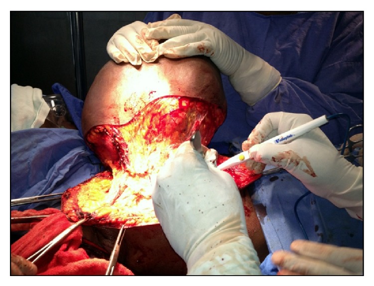
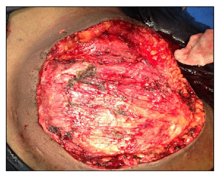
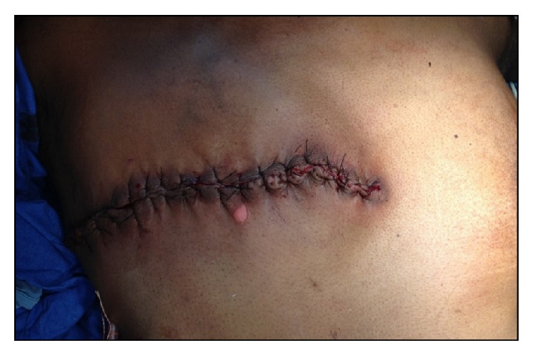
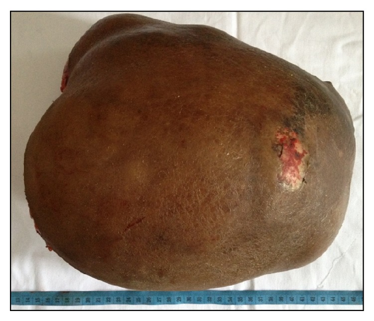
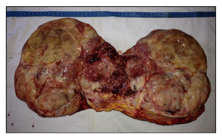
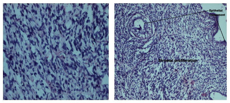
The term phyllodes tumour includes lesions ranging from completely benign tumours to malignant sarcomas. Clinically phyllodes tumours are smooth, rounded, and usually painless multinodular lesions indistinguishable from fibroadenomas. Percentage of phyllodes tumour classified as malignant ranges from 23% to 50%. We report a case of second largest phyllodes tumour in a 35-year-old lady who presented with swelling of right breast since 6 months, initially small in size, that progressed gradually to present size. Examination revealed mass in the right breast measuring 36×32 cms with lobulated firm surface and weighing 10 kgs. Fine needle aspiration cytology was reported as borderline phyllodes; however core biopsy examination showed biphasic neoplasm with malignant stromal component. Simple mastectomy was done and specimen was sent for histopathological examination which confirmed the core biopsy report. Postoperatively the patient received chemotherapy and radiotherapy. The patient is on follow-up for a year and has not shown any evidence of metastasis or recurrence.
Figures


Similar articles
-
The Enigma of Giant Phyllodes Tumour.Cureus. 2022 Mar 8;14(3):e22946. doi: 10.7759/cureus.22946. eCollection 2022 Mar. Cureus. 2022. PMID: 35411275 Free PMC article.
-
Massive Benign Phyllodes Tumour of Breast Complicating Pregnancy.J Clin Diagn Res. 2017 May;11(5):PD08-PD09. doi: 10.7860/JCDR/2017/26277.9929. Epub 2017 May 1. J Clin Diagn Res. 2017. PMID: 28658847 Free PMC article.
-
A large benign phyllodes tumour of the breast: A case report and literature review.Int J Surg Case Rep. 2017;39:192-195. doi: 10.1016/j.ijscr.2017.08.039. Epub 2017 Aug 23. Int J Surg Case Rep. 2017. PMID: 28854407 Free PMC article.
-
First case of transformation for breast fibroadenoma to high-grade malignant phyllodes tumor in an in vitro fertilization patient: misdiagnosis of recurrence, treatment and review of the literature.Eur Rev Med Pharmacol Sci. 2013 Sep;17(18):2495-8. Eur Rev Med Pharmacol Sci. 2013. PMID: 24089229 Review.
-
Fibroepithelial lesions; The WHO spectrum.Semin Diagn Pathol. 2017 Sep;34(5):438-452. doi: 10.1053/j.semdp.2017.05.006. Epub 2017 May 28. Semin Diagn Pathol. 2017. PMID: 28688536 Review.
Cited by
-
A Huge Benign Phyllodes Tumour of the Breast: a Rare Entity.Indian J Surg Oncol. 2019 Jun;10(2):389-391. doi: 10.1007/s13193-019-00911-y. Epub 2019 Mar 27. Indian J Surg Oncol. 2019. PMID: 31168269 Free PMC article. No abstract available.
-
Giant malignant phyllodes tumor with distant metastases: a case report and review of the literature.Hippokratia. 2022 Jan-Mar;26(1):41-45. Hippokratia. 2022. PMID: 37124283 Free PMC article.
-
Giant Benign Mammary Phyllodes Tumor: Report of a Case and Review of the Literature.Case Rep Oncol. 2021 Mar 1;14(1):123-133. doi: 10.1159/000510741. eCollection 2021 Jan-Apr. Case Rep Oncol. 2021. PMID: 33776693 Free PMC article. Review.
-
Phyllodes tumors of the breast in 2 sisters: Case report and review of literature.Medicine (Baltimore). 2017 Nov;96(46):e8552. doi: 10.1097/MD.0000000000008552. Medicine (Baltimore). 2017. PMID: 29145261 Free PMC article. Review.
References
-
- Salvadori B., Cusumano F., Del Bo R., Delledonne V., Grassi M., Rovini D., Saccozzi R., Andreola S., Clemente C. Surgical treatment of phyllodes tumors of the breast. Cancer. 1989;63(12):2532–2536. - PubMed
LinkOut - more resources
Full Text Sources
Other Literature Sources
