. 2014:2014:212096.
doi: 10.1155/2014/212096. Epub 2014 Dec 4.

Synthesis, characterization, and anticancer activity of new quinazoline derivatives against MCF-7 cells

Affiliations

Synthesis, characterization, and anticancer activity of new quinazoline derivatives against MCF-7 cells

Fadhil Lafta Faraj et al. ScientificWorldJournal. 2014.

Abstract

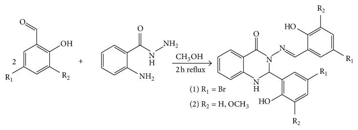
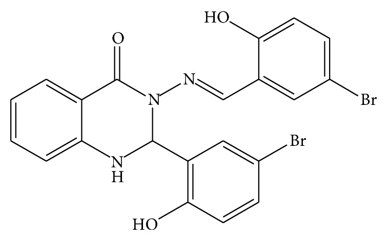
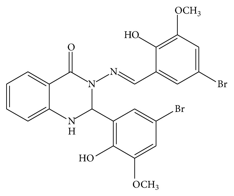
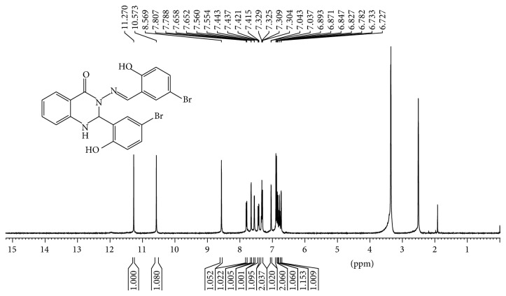
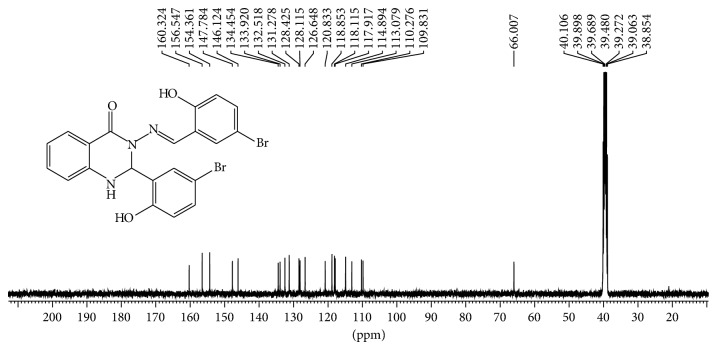
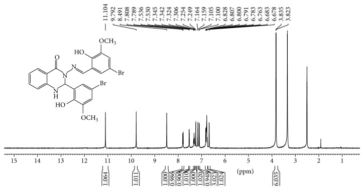
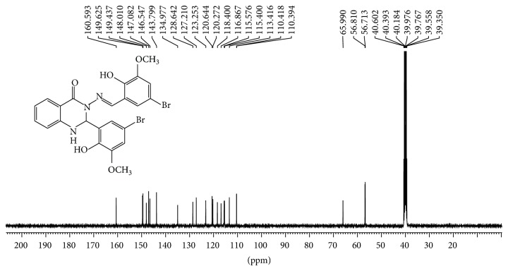
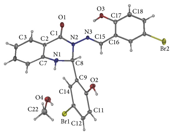
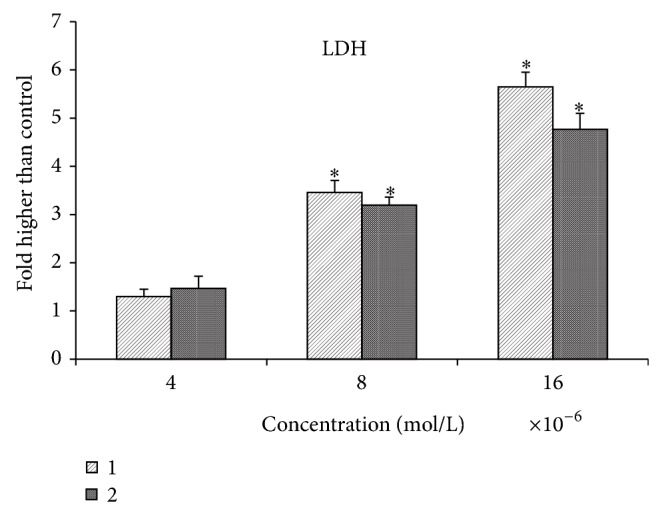
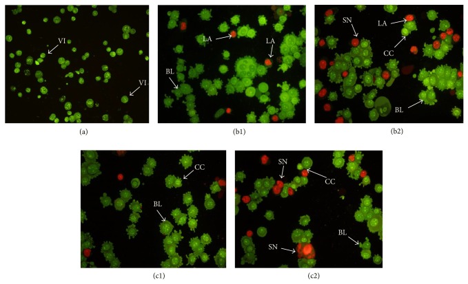
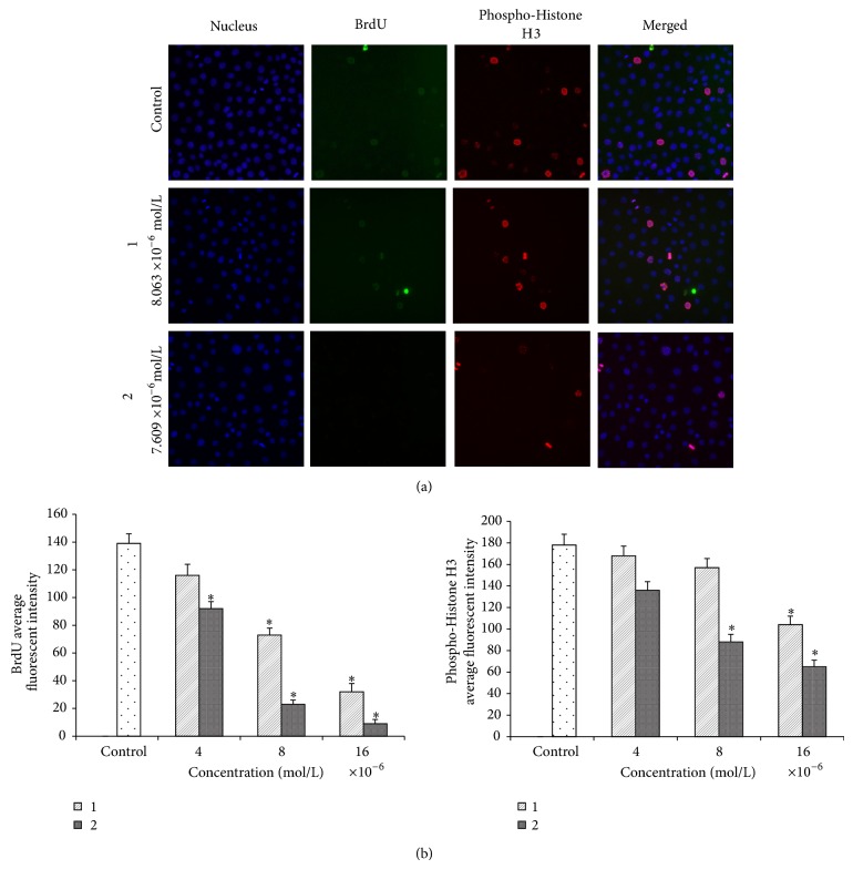
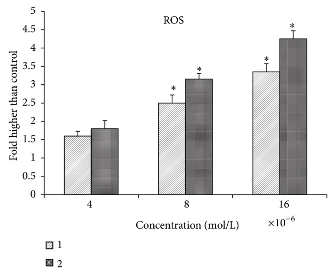
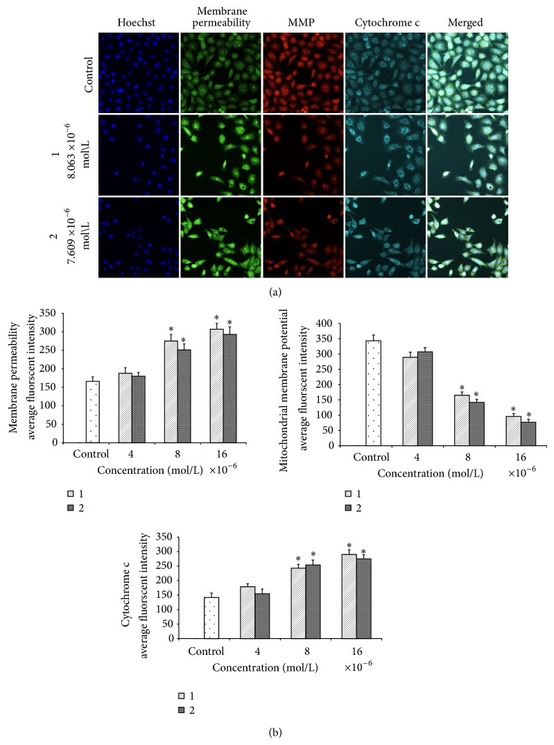
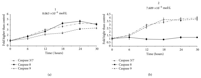
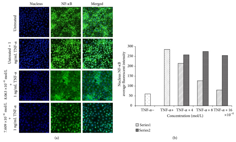
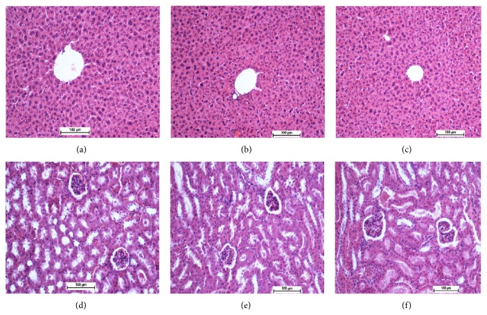
Two new synthesized and characterized quinazoline Schiff bases 1 and 2 were investigated for anticancer activity against MCF-7 human breast cancer cell line. Compounds 1 and 2 demonstrated a remarkable antiproliferative effect, with an IC50 value of 6.246×10(-6) mol/L and 5.910×10(-6) mol/L, respectively, after 72 hours of treatment. Most apoptosis morphological features in treated MCF-7 cells were observed by AO/PI staining. The results of cell cycle analysis indicate that compounds did not induce S and M phase arrest in cell after 24 hours of treatment. Furthermore, MCF-7 cells treated with 1 and 2 subjected to apoptosis death, as exhibited by perturbation of mitochondrial membrane potential and cytochrome c release as well as increase in ROS formation. We also found activation of caspases-3/7, -8, and -9 in compounds 1 and 2. Moreover, inhibition of NF-κB translocation in MCF-7 cells treated by compound 1 significantly exhibited the association of extrinsic apoptosis pathway. Acute toxicity results demonstrated the nontoxic nature of the compounds in mice. Our results showed significant activity towards MCF-7 cells via either intrinsic or extrinsic mitochondrial pathway and are potential candidate for further in vivo and clinical breast cancer studies.


Figures

Figure 1
Figure 1
Quinazoline-4-one.
Figure 2
Figure 2
The reaction pathway for the synthesis of compounds (1) and (2).
Figure 3
Figure 3
Compound (1).
Figure 4
Figure 4
Compound (2).
Figure 5
Figure 5
1H NMR spectrum of compound (1) in (400 MHz, DMSO-d6).
Figure 6
Figure 6
13C NMR spectrum of compound (1) in (100 MHz, DMSO-d6).
Figure 7
Figure 7
1H NMR spectrum of a compound (2) in (400 MHz, DMSO-d6).
Figure 8
Figure 8
13C NMR spectrum of compound (2) in (100 MHz, DMSO-d6).
Figure 9
Figure 9
Crystal structure of compound (1).
Figure 10
Figure 10
The LDH release assay revealed the significant cytotoxicity of the quinazolinone-based compound on MCF-7 cells at concentrations 4 × 10⁻⁶, 8 × 10⁻⁶, and 16 × 10−6 mol/L.
Figure 11
Figure 11
Fluorescent micrographs of AO/PI-double-stained MCF-7 cells. (a) Untreated MCF-7 cells exhibit normal structures. (b1) and (c1) Early apoptosis features, namely, blebbing and chromatin condensation as well as late apoptotic cells, were detected after 24 h of treatment with (1) and (2). (b2) and (c2) Late apoptosis and secondary necrosis were obsereved after 48 h of treatment with (1) and (2), respectively (magnification: 200x). VI: viable cells; CC: chromatin condensation; BL: blebbing of the cell membrane; LA: late apoptosis; SN: secondary necrosis.
Figure 12
Figure 12
Cell cycle analysis. (a) Effect of (1) and (2) on cell cycle arrest. After incubation with DMSO or different concentrations of (1) and (2) for 24 h, MCF-7 cells were stained with BrdU and phosphohistone H3 and subjected to the Cellomics ArrayScan HCS reader for cell cycle analysis. (b) Representative bar charts indicating that treatment of both compounds markedly decreased BrdU and phosphohistone H3 fluorescence intensities in treated MCF-7 cells. Data were expressed as the mean ± SD of fluorescence intensity readings for three independent experiments.
Figure 13
Figure 13
Effect of MMD compound on the generation of ROS. The level of ROS significantly elevated at 8 × 10⁻⁶ and 16 × 10−6 mol/L concentrations.
Figure 14
Figure 14
Effects of the Quinazoline Schiff bases on nuclear morphology, membrane permeability, mitochondrial membrane potential (MMP), and cytochrome c release. (a) Representative images of MCF-7 cells treated with medium alone and at 4 × 10⁻⁶, 8 × 10⁻⁶, and 16 × 10−6 mol/L concentrations of compounds and stained with Hoechst 33342 for nuclear, cytochrome c, membrane permeability, and MMP dyes. Both compound induced a noteworthy elevation in membrane permeability and cytochrome c release and a marked reduction in mitochondrial membrane potential (magnification: 200x). (b) Representative bar charts indicating dose-dependent increased cell permeability reduced MMP and increased cytochrome c release in treated MCF-7 cells.
Figure 15
Figure 15
Relative luminescence time-dependent expression of caspases-3/7, -8, and -9 in MCF-7 cells treated with (1) and (2) at concentrations of 8 × 10⁻⁶ mol/L and 7.6 × 10⁻⁶ mol/L, respectively, after 24 hours incubation.
Figure 16
Figure 16
NF-κB translocation. (a) Photographs of the intracellular targets of stained MCF-7 cells that were exposed to (1) and (2) at concentrations of 8 × 10⁻⁶ mol/L and 7.6 × 10⁻⁶ mol/L, respectively, for 3 hours and then stimulated for 30 minutes with 1 ng/mL TNF-α (NF-κB activation). (b) Representative bar chart showing translocation of TNF-α-induced NF-κB nuclear in MCF-7 cells for different concenataions.
Figure 17
Figure 17
Histological sections in acute toxicity test (H&E staining, 20x). Histological sections of liver (first row) and kidney (second row). Untreated mices (control group) received 5 mL/kg vehicle (5% Tween 20) ((a) and (d)). Animals treated with 250 mg/kg are ((b) and (e)) and ((c) and (f)) for compounds 1 and 2, respectively. There are no significant differences in structures of liver and kidney between treated and control group.

References

    1. Saurav K., Garima M., Pradeep S., Jha K. K., Khosa R. L., Gupta S. K. Quinazoline-4-one: a highly important hetrocycle with diverse biological activities. Der Chemica Sinica. 2011;2(4):36–58.
    1. Selvam T. P., Kumar P. V., Vijayaraj P. Quinazoline marketed drugs—a review. Research in Pharmacy. 2011;1(1):1–21.
    1. A. Shetha, I. A. Wijdan Synthesis and characterization of new quinazoline-4(3H)-one Schiff bases. Journal of Chemical and Pharmaceutical Research. 2013;5(7):42–45.
    1. Vagdevi H. M., Lokesh M. R., Gowdarshivannanavar B. C. Synthesis and antioxidant activity of 3-substituted Schiff bases of quinazoline-2,4-diones. International Journal of ChemTech Research. 2012;4(4):1527–1533.
    1. Krishnan S. K., Ganguly S., Veerasamy R., Jan B. Synthesis, antiviral and cytotoxic investigation of 2-phenyl-3-substituted quinazolin-4(3H)-ones. European Review for Medical and Pharmacological Sciences. 2011;15(6):673–681. - PubMed

Publication types

MeSH terms

LinkOut - more resources