. 2014 Dec 30;9(12):e116295.
doi: 10.1371/journal.pone.0116295. eCollection 2014.

Inflammatory micro-environmental cues of human atherothrombotic arteries confer to vascular smooth muscle cells the capacity to trigger lymphoid neogenesis

Affiliations

Inflammatory micro-environmental cues of human atherothrombotic arteries confer to vascular smooth muscle cells the capacity to trigger lymphoid neogenesis

Kevin Guedj et al. PLoS One. .

Abstract

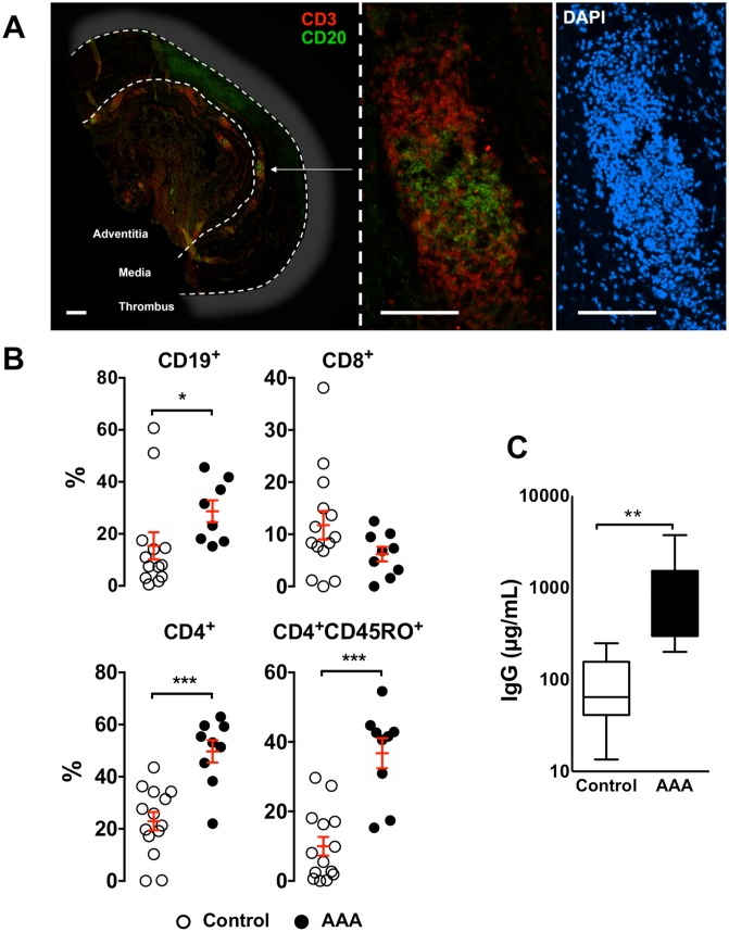
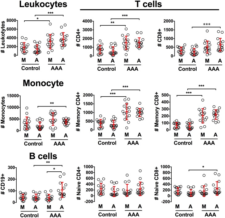
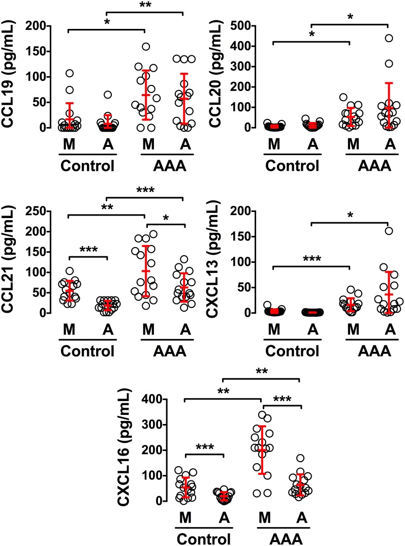
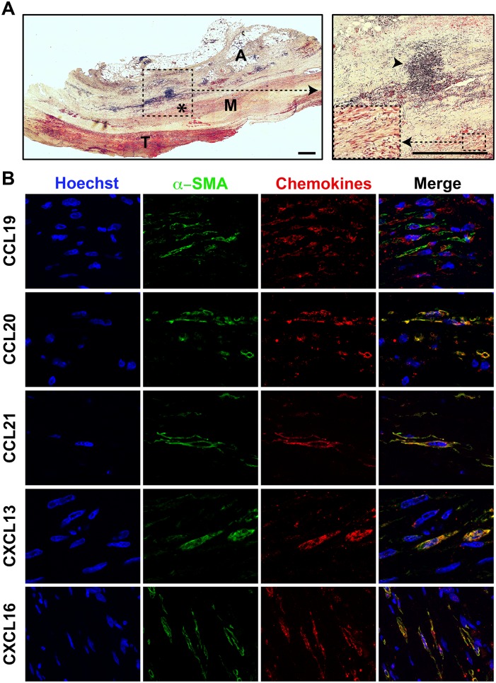
Background: Experimental atherosclerosis is characterized by the formation of tertiary lymphoid structures (TLOs) within the adventitial layer, which involves the chemokine-expressing aortic smooth muscle cells (SMCs). TLOs have also been described around human atherothrombotic arteries but the mechanisms of their formation remain poorly investigated. Herein, we tested whether human vascular SMCs play the role of chemokine-expressing cells that would trigger the formation of TLOs in atherothrombotic arteries.

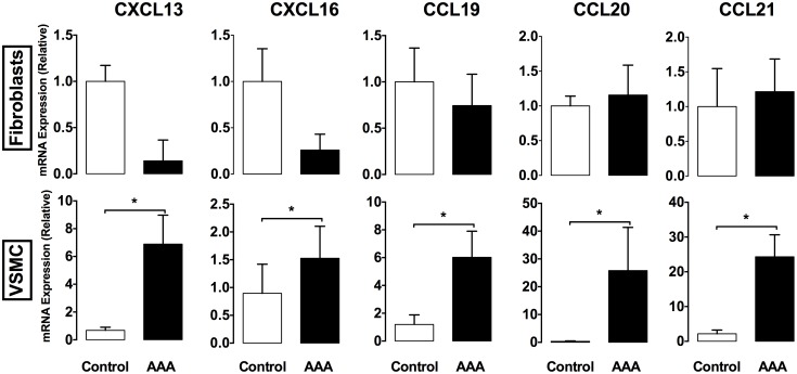
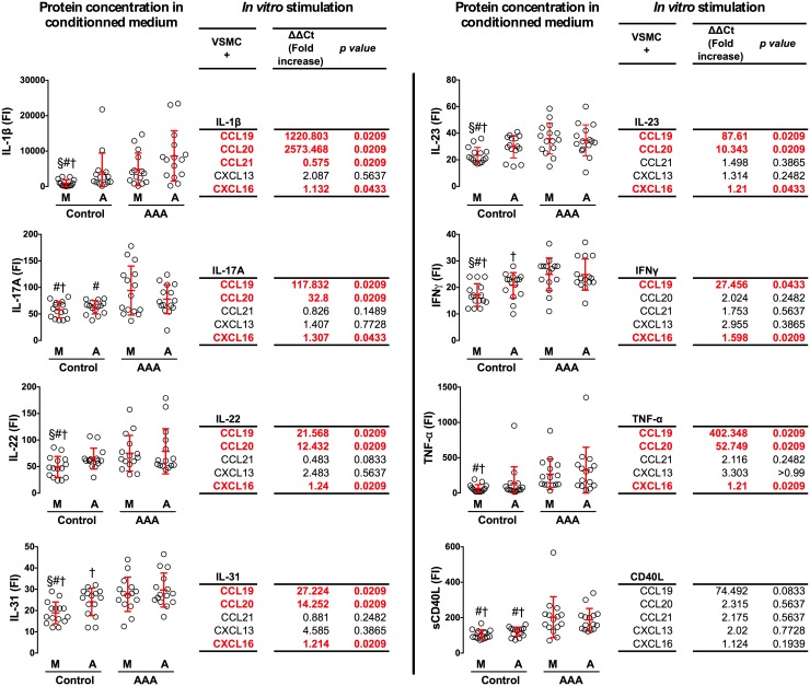
Results: We first characterized, by flow cytometry and immunofluorescence analysis, the prevalence and cell composition of TLOs in human abdominal aneurysms of the aorta (AAAs), an evolutive form of atherothrombosis. Chemotaxis experiments revealed that the conditioned medium from AAA tissues recruited significantly more B and T lymphocytes than the conditioned medium from control (N-AAA) tissues. This was associated with an increase in the concentration of CXCL13, CXCL16, CCL19, CCL20, and CCL21 chemokines in the conditioned medium from AAA tissues. Immunofluorescence analysis of AAA cryosections revealed that α-SMA-positive SMCs were the main contributors to the chemokine production. These results were confirmed by RT-qPCR assays where we found that primary vascular SMCs from AAA tissues expressed significantly more chemokines than SMCs from N-AAA. Finally, in vitro experiments demonstrated that the inflammatory cytokines found to be increased in the conditioned medium from AAA were able to trigger the production of chemokines by primary SMCs.

Conclusion: Together, these results suggest that human vascular SMCs in atherothrombotic arteries, in response to inflammatory signals, are converted into chemokine-expressing cells that trigger the recruitment of immune cells and the formation of aortic TLOs.


Conflict of interest statement

Competing Interests: The authors have declared that no competing interests exist.

Figures

Figure 1
Figure 1. Organized Tertiary Lymphoid Structures in human Abdominal Aortic Aneurysm.
(A) Representative immunohistochemical staining of CD3+ T cells (red), CD20+ B cells (green) and nuclei (DAPI, blue) performed on human AAA cross-sections (scale bar = 1 mm). (B) Flow cytometry analysis of the percentage of CD19+ B cells, total CD8+, CD4+and memory (CD4+CD45RO+) T cells in adventitia from AAAs (n = 9) or control aortas (n = 14). (C) Box plot showing the total IgG levels in conditioned medium prepared from adventitial tissues of AAAs (n = 14) and control aortas (n = 5) quantified by ELISA. *, p<0.05; **, p<0.01; ***, p<0.001.
Figure 2
Figure 2. The aneurysmal aortic tissue promotes the chemoattraction of immune cells.
Chemotaxis assay on conditioned medium prepared from the media (M) and adventitia (A) of AAAs (n = 15) and control aortas (n = 15). Plotted is the number of total leukocytes, monocytes, CD19+ B cells, CD44+CD62L memory CD4+ or CD8+ T cells, CD44CD62L+ naïve CD4+ or CD8+ T cells that have migrated toward the well containing the conditioned medium. *, p<0.05; **, p<0.01; ***, p<0.001.
Figure 3
Figure 3. Increased concentration of chemokines in conditioned medium from AAA tissues.
The concentration of CCL19, CCL20, CCL21, CXCL13 and CXCL16 was assessed by ELISA assays on conditioned medium prepared from the media (M) and adventitia (A) of AAAs (n = 15) and control aortas (n = 15). *, p<0.05; **, p<0.01; ***, p<0.001.
Figure 4
Figure 4. VSMCs in the vicinity of human aortic TLOs produce chemokines.
(A) Masson’s trichrome staining performed on a human AAA cross-section. The dashed square indicates the area that is magnified on the right inset. The asterisk represents the smooth muscle cell-rich area that is magnified in the dashed square box in the right inset (M = media; A = adventitia; SLO = Secondary Lymphoid Organ; scale bar = 2 mm). (B) Representative immunohistochemical staining of α-SMA+ VSMCs (green), CCL19, CCL20, CCL21, CXCL13 and CXCL16 chemokines (red) and nuclei (DAPI, blue) performed on human AAA cryosections (scale bar = 50 µm; n = 5).
Figure 5
Figure 5. VSMCs, but not fibroblasts, from AAA patients express chemokines.
(A) The expression of CCL19, CCL20, CCL21, CXCL13 and CXCL16 chemokines was determined by RT-qPCR on RNA extracted from primary cultures of adventitial fibroblasts and medial VSMCs from AAA and healthy subjects. Data were analyzed using the 2−ΔΔCt Pfaffl formula in which data from AAAs were compared to Ct from control aortas, normalized to the Ct values of the β-actin. Data are representative of 4 independent experiments. *, p<0.05.
Figure 6
Figure 6. Inflammatory chemokines produced in AAA tissues trigger the expression of chemokines by VSMC.
The graphs represent the relative concentrations of IL-1β, IL-17A, IL-22, IL-31, IL-23, IFNγ, TNF-α and CD40L that were assessed by ELISA assays on conditioned medium prepared from the media (M) and adventitia (A) of AAAs (n = 15) and control aortas (n = 15; §, #, †, p<0.05 vs Control A, AAA M and AAA A, respectively). The tables represent the expression of CCL19, CCL20, CCL21, CXCL13, and CXCL16 chemokines by human VSMCs that were stimulated for 15 hours with rIL-1β (5 ng/mL), rIL-17A (50 ng/mL), rIL-22 (10 ng/mL), rIL-31 (50 ng/mL), rIL-23 (25 ng/mL), rIFNγ (10 ng/mL), rTNF-α (1 ng/mL), and rCD40L (100 ng/mL). These cytokine concentrations were 2 log higher than those detected in the conditioned medium, so as to compensate for the in vivo situation where cells are subjected to chronic, additive and combined stimulations. The relative expression was determined by RT-qPCR on extracted RNA, and data were analyzed using the 2−ΔΔCt Pfaffl formula in which Ct values from stimulated VSMCs were compared to unstimulated cells, and normalized to the Ct values of β-actin. Data are representative of 4 independent experiments.

References

    1. Hansson GK (2001) Immune mechanisms in atherosclerosis. Arterioscler Thromb Vasc Biol 21:1876–1890. - PubMed
    1. Aloisi F, Pujol-Borrell R (2006) Lymphoid neogenesis in chronic inflammatory diseases. Nat Rev Immunol 6:205–217. - PubMed
    1. Houtkamp MA, de Boer OJ, van der Loos CM, van der Wal AC, Becker AE (2001) Adventitial infiltrates associated with advanced atherosclerotic plaques: structural organization suggests generation of local humoral immune responses. J Pathol 193:263–269. - PubMed
    1. Grabner R, Lotzer K, Dopping S, Hildner M, Radke D, et al. (2009) Lymphotoxin beta receptor signaling promotes tertiary lymphoid organogenesis in the aorta adventitia of aged ApoE−/− mice. J Exp Med 206:233–248. - PMC - PubMed
    1. Dutertre CA, Clement M, Morvan M, Schakel K, Castier Y, et al. (2014) Deciphering the stromal and hematopoietic cell network of the adventitia from non-aneurysmal and aneurysmal human aorta. PLoS One 9:e89983. - PMC - PubMed

Publication types

MeSH terms

LinkOut - more resources