Review
. 2014 Dec 16;20 Suppl 1(Suppl 1):S43-58.
doi: 10.2119/molmed.2014.00232.

An expanding role for interleukin-1 blockade from gout to cancer

Affiliations
Review

An expanding role for interleukin-1 blockade from gout to cancer

Charles Anthony Dinarello. Mol Med. .

Abstract

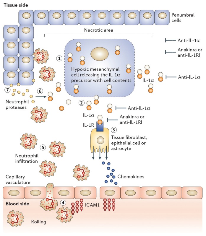
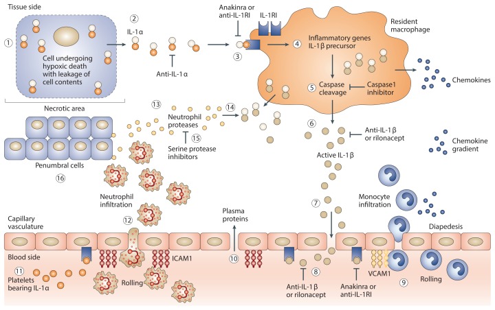
There is an expanding role for interleukin (IL)-1 in diseases from gout to cancer. More than any other cytokine family, the IL-1 family is closely linked to innate inflammatory and immune responses. This linkage is because the cytoplasmic segment of all members of the IL-1 family of receptors contains a domain, which is highly homologous to the cytoplasmic domains of all toll-like receptors (TLRs). This domain, termed "toll IL-1 receptor (TIR) domain," signals as does the IL-1 receptors; therefore, inflammation due to the TLR and the IL-1 families is nearly the same. Fundamental responses such as the induction of cyclo-oxygenase type 2, increased surface expression of cellular adhesion molecules and increased gene expression of a broad number of inflammatory molecules characterizes IL-1 signal transduction as it does for TLR agonists. IL-1β is the most studied member of the IL-1 family because of its role in mediating autoinflammatory disease. However, a role for IL-1α in disease is being validated because of the availability of a neutralizing monoclonal antibody to human IL-1α. There are presently three approved therapies for blocking IL-1 activity. Anakinra is a recombinant form of the naturally occurring IL-1 receptor antagonist, which binds to the IL-1 receptor and prevents the binding of IL-1β as well as IL-1α. Rilonacept is a soluble decoy receptor that neutralizes primarily IL-1β but also IL-1α. Canakinumab is a human monoclonal antibody that neutralizes only IL-1β. Thus, a causal or significant contributing role can be established for IL-1β and IL-1α in human disease.


Figures

Figure 1
Figure 1
Initiation of sterile inflammation by IL-1α following an ischemic event. ①, In the necrotic area, dying cells lose membrane integrity. ②, Dying cells release their contents including the IL-1α precursor. Anti–IL-1α antibodies neutralize IL-1α at this step. ③, IL-1α binds to IL-1R type I (IL-1RI) on nearby resident fibroblasts, epithelial cells or in the brain astrocytes, releasing chemokines and establishing a chemokine gradient. Anakinra or anti–IL-1RI prevents this step. The chemokine gradient facilitates the passage to blood neutrophils into the ischemic area. ④, Capillaries in the ischemic tissues express the intracellular adhesion molecule-1 (ICAM-1). Circulating blood neutrophils roll on the endothelium, adhere to ICAM-1 and enter the ischemic tissue via diapedesis. ⑤, The number of neutrophils increases into the area of the necrotic event; the presence of local IL-1 prolongs the survival of neutrophils at this step. ⑥, Neutrophil proteases cleave the extracellular IL-1α precursor into mature, more active forms. ⑦, Neutrophils scavenge dying cells and release proteases that contribute to the destruction of penumbral cells. Image reproduced (no permission needed) from Dinarello et al. (137): Charles A Dinarello, Anna Simon, Jos W M van der Meer. Treating inflammation by blocking interleukin-1 in a broad spectrum of diseases. Nature Reviews Drug Discovery. 2012;11:633–52.
Figure 2
Figure 2
Role of IL 1β in sterile inflammation. ①, Following an ischemic event, cells undergo hypoxic damage, lose membrane integrity, and the dying cell releases cell content (see Figure 1). ②, The preformed IL-1α precursor is released. Anti–IL-1α antibodies neutralize IL-1α at this step. ③, IL-1α binds to IL-1 receptor type I (IL-1RI) on nearby resident macrophages. Anakinra or anti–IL-1RI antibodies prevent IL-1α activity at this step. ④, Triggered by IL-1α binding to the IL-1RI, resident macrophages synthesize inflammatory genes as well as the IL-1β precursor. ⑤, Intracellular processing of the IL-1β precursor by caspase-1. Caspase-1 inhibitors prevent the processing of IL-1β. ⑥, Active secretion of active IL-1β. Antibodies to IL-1β or rilonacept neutralize IL-1β in the extracellular compartment at this step. ⑦, With breakdown of the vascular integrity in the necrotic area, IL-1β gains access to the vascular compartment. ⑧, IL-1β binds to IL-1RI on capillaries and induces vascular cell adhesion molecule-1 (VCAM-1). ⑨, Blood monocytes roll along the endothelium and bind to VCAM-1, followed by emigration into the ischemic tissue via diapedesis. Increasing numbers of monocytes become a source of greater production of IL-1β. ⑩, Opening of the endothelial junction resulting in capillary leak with the passage of plasma proteins into the ischemic area. ⑪, Platelet-derived IL-1α binds to the endothelial IL-1RI and induces ICAM-1. ⑫, Large numbers of neutrophils enter the tissue space, and the presence of local IL-1 prolongs the survival of neutrophils. ⑬, Release of neutrophil proteases. ⑭, IL-1β precursor released into the extracellular space is cleaved by serine proteases generating active IL-1β. Natural inhibitors of serine proteases such as α1-antitrypsin prevent the extracellular processing of the IL-1β precursor. ⑮, Increasing numbers of neutrophils surround the necrotic area, scavenging dead cells and debris. ⑯, Damaging neutrophilic proteases attack and injure penumbral cells, increasing the loss of function. Blocking IL-1 cannot restore the necrotic tissue but reduces the loss of the penumbral cells. Image modified (no permission needed) from Dinarello et al. (137): Charles A Dinarello, Anna Simon, Jos W M van der Meer. Treating inflammation by blocking interleukin-1 in a broad spectrum of diseases. Nature Reviews Drug Discovery. 2012;11:633–52.
None

References

    1. Dinarello CA, Renfer L, Wolff SM. Human leukocytic pyrogen: purification and development of a radioimmunoassay. Proc Natl Acad Sci U S A. 1977;74:4624–7. - PMC - PubMed
    1. Murphy PA, Chesney PJ, Wood WB., Jr Further purification of rabbit leukocyte pyrogen. J Lab Clin Med. 1974;83:310–22. - PubMed
    1. Kawakami M, Pekala PH, Lane MD, Cerami A. Lipoprotein lipase suppression in 3T3-L1 cells by an endotoxin-induced mediator from exudate cells. Proc Natl Acad Sci U S A. 1982;79:912–6. - PMC - PubMed
    1. Dinarello CA, et al. Tumor necrosis factor (cachectin) is an endogenous pyrogen and induces production of interleukin 1. J Exp Med. 1986;163:1433–50. - PMC - PubMed
    1. Dinarello CA. Interleukin-1 and the pathogenesis of the acute-phase response. N Engl J Med. 1984;311:1413–8. - PubMed

Publication types