

Genome-wide identification and comprehensive analyses of the kinomes in four pathogenic microsporidia species
- PMID: 25549259
- PMCID: PMC4280135
- DOI: 10.1371/journal.pone.0115890
Genome-wide identification and comprehensive analyses of the kinomes in four pathogenic microsporidia species
Abstract
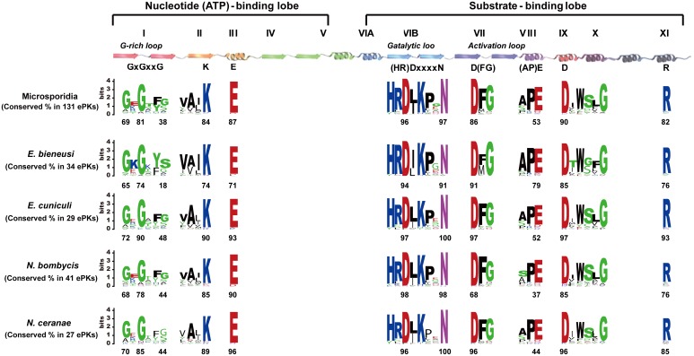
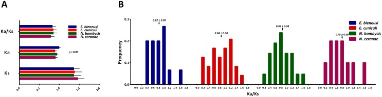
Microsporidia have attracted considerable attention because they infect a wide range of hosts, from invertebrates to vertebrates, and cause serious human diseases and major economic losses in the livestock industry. There are no prospective drugs to counteract this pathogen. Eukaryotic protein kinases (ePKs) play a central role in regulating many essential cellular processes and are therefore potential drug targets. In this study, a comprehensive summary and comparative analysis of the protein kinases in four microsporidia—Enterocytozoon bieneusi, Encephalitozoon cuniculi, Nosema bombycis and Nosema ceranae—was performed. The results show that there are 34 ePKs and 4 atypical protein kinases (aPKs) in E. bieneusi, 29 ePKs and 6 aPKs in E. cuniculi, 41 ePKs and 5 aPKs in N. bombycis, and 27 ePKs and 4 aPKs in N. ceranae. These data support the previous conclusion that the microsporidian kinome is the smallest eukaryotic kinome. Microsporidian kinomes contain only serine-threonine kinases and do not contain receptor-like and tyrosine kinases. Many of the kinases related to nutrient and energy signaling and the stress response have been lost in microsporidian kinomes. However, cell cycle-, development- and growth-related kinases, which are important to parasites, are well conserved. This reduction of the microsporidian kinome is in good agreement with genome compaction, but kinome density is negatively correlated with proteome size. Furthermore, the protein kinases in each microsporidian genome are under strong purifying selection pressure. No remarkable differences in kinase family classification, domain features, gain and/or loss, and selective pressure were observed in these four species. Although microsporidia adapt to different host types, the coevolution of microsporidia and their hosts was not clearly reflected in the protein kinases. Overall, this study enriches and updates the microsporidian protein kinase database and may provide valuable information and candidate targets for the design of treatments for pathogenic diseases.
Conflict of interest statement
Figures


References
-
- Bhat SA, Bashir I, Kamili AS (2009) Microsporidiosis of silkworm, Bombyx mori L.(Lepidoptera-Bombycidae): a review. Afr J Agric Res 4:1519–1523.
-
- Chen Y, Evans JD, Smith IB, Pettis JS (2008) Nosema ceranae is a long-present and wide-spread microsporidian infection of the European honey bee (Apis mellifera) in the United States. Journal of Invertebrate Pathology 97:186–188. - PubMed
-
- Cox-Foster DL, Conlan S, Holmes EC, Palacios G, Evans JD, et al. (2007) A metagenomic survey of microbes in honey bee colony collapse disorder. Science 318:283–287. - PubMed
-
- Higes M, Martín-Hernández R, Botías C, Bailón EG, González-Porto AV, et al. (2008) How natural infection by Nosema ceranae causes honeybee colony collapse. Environmental Microbiology 10:2659–2669. - PubMed
Publication types
MeSH terms
Substances
LinkOut - more resources
Full Text Sources
Other Literature Sources
