. 2014 Dec 30;9(12):e116003.
doi: 10.1371/journal.pone.0116003. eCollection 2014.

Proteomic screening for amyloid proteins

Affiliations

Proteomic screening for amyloid proteins

Anton A Nizhnikov et al. PLoS One. .

Abstract

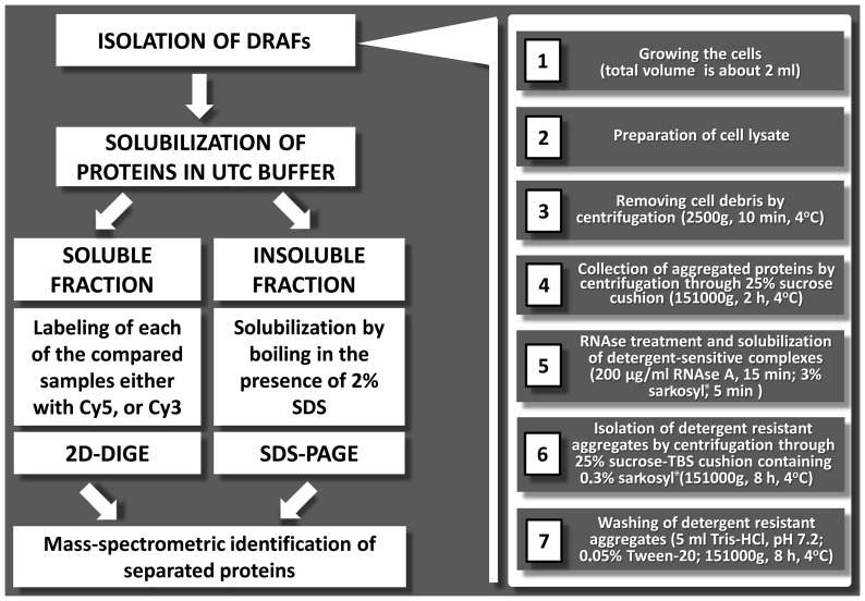
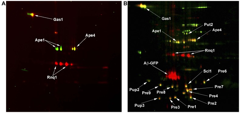
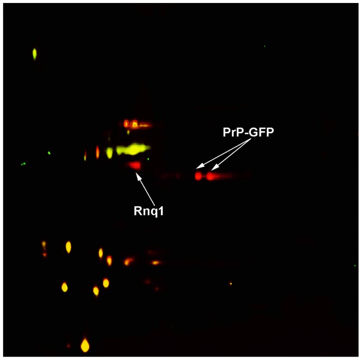
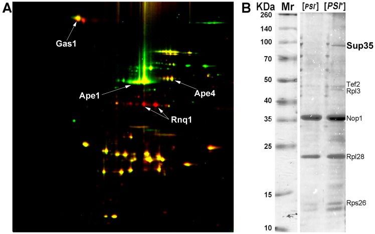
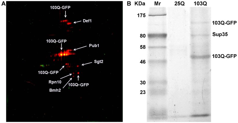
Despite extensive study, progress in elucidation of biological functions of amyloids and their role in pathology is largely restrained due to the lack of universal and reliable biochemical methods for their discovery. All biochemical methods developed so far allowed only identification of glutamine/asparagine-rich amyloid-forming proteins or proteins comprising amyloids that form large deposits. In this article we present a proteomic approach which may enable identification of a broad range of amyloid-forming proteins independently of specific features of their sequences or levels of expression. This approach is based on the isolation of protein fractions enriched with amyloid aggregates via sedimentation by ultracentrifugation in the presence of strong ionic detergents, such as sarkosyl or SDS. Sedimented proteins are then separated either by 2D difference gel electrophoresis or by SDS-PAGE, if they are insoluble in the buffer used for 2D difference gel electrophoresis, after which they are identified by mass-spectrometry. We validated this approach by detection of known yeast prions and mammalian proteins with established capacity for amyloid formation and also revealed yeast proteins forming detergent-insoluble aggregates in the presence of human huntingtin with expanded polyglutamine domain. Notably, with one exception, all these proteins contained glutamine/asparagine-rich stretches suggesting that their aggregates arose due to polymerization cross-seeding by human huntingtin. Importantly, though the approach was developed in a yeast model, it can easily be applied to any organism thus representing an efficient and universal tool for screening for amyloid proteins.


Conflict of interest statement

Competing Interests: The authors have declared that no competing interests exist.

Figures

Figure 1
Figure 1. Schematic representation of PSIA.
Left panel illustrates the main stages of PSIA. Right panel describes the procedures for DRAF isolation. For details, see Materials and methods. *1% and 0.1% SDS can be used instead of 3% and 0.3% sarkosyl in steps 5 and 6, respectively.
Figure 2
Figure 2. 2D-DIGE image of proteins of SDS- (A) or sarkosyl-insoluble (B) aggregates isolated from the BY4742 [PIN +] strain expressing Aβ-GFP and from its [pin ] derivative expressing non-fused GFP.
Spots corresponding to proteins from [PIN +] cells expressing Aβ-GFP are red, while proteins from GFP-containing cells (control) are green. Yellow spots correspond to proteins present in both compared samples. Proteins identified by mass-spectrometry are indicated. Identification data are presented in Table 1 (SDS-insoluble aggregates) and 2 and 3(sarkosyl-insoluble aggregates). Mass-spectra of identified proteins are listed in S1–S3 Tables, correspondingly.
Figure 3
Figure 3. 2D-DIGE image of proteins of sarkosyl-insoluble aggregates isolated from the BY4742 [PIN +] strain expressing PrP-GFP and from its [pin ] derivative expressing non-fused GFP.
Spots corresponding to proteins from [PIN +] cells expressing PrP-GFP are red, while those from GFP-containing cells (control) are green. Yellow spots correspond to proteins present in both compared samples. Identification data are presented in Table 2; mass spectra are shown in S2 Table.
Figure 4
Figure 4. 2D-DIGE image of proteins of sarkosyl-insoluble aggregates isolated from the GT81-1C [PSI +][PIN +] strain and its [psi ][pin ] derivative (A).
Spots corresponding to proteins from [PSI +][PIN +] lysate and the [psi][pin ] control are red and green, respectively, while yellow spots correspond to proteins present in both compared samples. SDS-PAGE image of proteins from the same DRAFs that were insoluble in UTC buffer (B). Proteins identified by mass-spectrometry are indicated. Mr – protein molecular mass markers. Identification data are presented in Table 4; mass spectra are shown in S4 Table.
Figure 5
Figure 5. Identification of proteins forming SDS-insoluble aggregates in cells expressing 103Q-GFP.
2D-DIGE image of proteins of aggregates isolated using SDS (A). Spots corresponding to proteins from cells expressing 103Q-GFP and 25Q-GFP (control) are red and green, respectively. SDS-PAGE image of proteins from the same DRAFs that were insoluble in UTC buffer (B). Mr – protein molecular mass markers. Proteins identified by mass-spectrometry are indicated. Identification data are presented in Table 5 and S5 Table.

References

    1. Sipe JD, Benson MD, Buxbaum JN, Ikeda S, Merlini G, et al. (2012) Amyloid fibril protein nomenclature: 2012 recommendations from the Nomenclature Committee of the International Society of Amyloidosis. Amyloid 19:167–70. - PubMed
    1. Moreno-Gonzalez I, Soto C (2011) Misfolded protein aggregates: mechanisms, structures and potential for disease transmission. Semin Cell Dev Biol 22:482–7. - PMC - PubMed
    1. Bhak G, Choe Y, Paik S (2009) Mechanism of amyloidogenesis: nucleation-dependent fibrillation versus double-concerted fibrillation. BMB Reports 42:541–51. - PubMed
    1. Prusiner SB (1982) Novel proteinaceous infections particles cause scrapie. Science 216:136–44. - PubMed
    1. Wickner RB (1994) [URE3] as an altered Ure2 protein: evidence for a prion analog in Saccharomyces cerevisiae . Science 264:566–69. - PubMed

Publication types

MeSH terms

LinkOut - more resources