. 2014 Dec 30;9(12):e115720.
doi: 10.1371/journal.pone.0115720. eCollection 2014.

CCR7 expressing mesenchymal stem cells potently inhibit graft-versus-host disease by spoiling the fourth supplemental Billingham's tenet

Affiliations

CCR7 expressing mesenchymal stem cells potently inhibit graft-versus-host disease by spoiling the fourth supplemental Billingham's tenet

Hong Li et al. PLoS One. .

Abstract

The clinical acute graft-versus-host disease (GvHD)-therapy of mesenchymal stem cells (MSCs) is not as satisfactory as expected. Secondary lymphoid organs (SLOs) are the major niches serve to initiate immune responses or induce tolerance. Our previous study showed that CCR7 guide murine MSC line C3H10T1/2 migrating to SLOs. In this study, CCR7 gene was engineered into murine MSCs by lentivirus transfection system (MSCs/CCR7). The immunomodulatory mechanism of MSCs/CCR7 was further investigated. Provoked by inflammatory cytokines, MSCs/CCR7 increased the secretion of nitric oxide and calmed down the T cell immune response in vitro. Immunofluorescent staining results showed that transfused MSCs/CCR7 can migrate to and relocate at the appropriate T cell-rich zones within SLOs in vivo. MSCs/CCR7 displayed enhanced effect in prolonging the survival and alleviating the clinical scores of the GvHD mice than normal MSCs. Owing to the critical relocation sites, MSCs/CCR7 co-infusion potently made the T cells in SLOs more naïve like, thus control T cells trafficking from SLOs to the target organs. Through spoiling the fourth supplemental Billingham's tenet, MSCs/CCR7 potently inhibited the development of GvHD. The study here provides a novel therapeutic strategy of MSCs/CCR7 infusion at a low dosage to give potent immunomodulatory effect for clinical immune disease therapy.


Conflict of interest statement

Competing Interests: The authors have declared that no competing interests exist.

Figures

Figure 1
Figure 1. CCR7 expression in murine and human MSCs.
(A) RT-PCR analysis of CCR7 mRNA expression level in the human primary MSCs derived from bone marrow (hBM-MSCs) and umbilical cord (hUC-MSCs), human peripheral blood cells (hPBC) served as positive control. Human housekeeping gene of GAPDH acted as internal standard control. (B) FCM analysis of CCR7 protein on the cell surface of human BM-MSCs and UC-MSCs. (C)RT-PCR analysis of CCR7 mRNA level in the primary MSCs, splenic mononucleocytes (SPC) served as positive controls. Murine housekeeping gene of GAPDH acted as internal standards control. (D)FCM analysis of CCR7 protein on the cell surface of SPC and murine MSCs derived from BALB/c and C57BL/6 mice. (E) Murine CCR7 gene was engineered into murine MSCs by lentiviral vector transduction. RT-PCR analysis of CCR7 and eGFP mRNA expression in the MSCs, MSCs/eGFP and MSCs/CCR7. (F) FCM analysis of CCR7 protein on the cell surface of the MSCs, MSCs/eGFP and MSCs/CCR7. Data are representative of three independent experiments.
Figure 2
Figure 2. iNOS expression in MSCs/CCR7 is elicit by inflammatory cytokines.
(A) iNOS expression was examined by real-time PCR in the MSCs, MSCs/eGFP and MSCs/CCR7 without or with IFNγ plus TNFα treatment. (B) iNOS expression was examined by in situ immunofluorescence staining. (C) MSCs, MSCs/eGFP and MSCs/CCR7 were stimulated with IFNγ plus TNFα. NO in the supernatant was examined by Griess assay. Data shown are mean±S.D. of a representative of 3 independent experiments. **P<0.01 and ***P<0.001 when compared with the cells without cytokine stimulation.
Figure 3
Figure 3. MSCs/CCR7 inhibit T cells proliferation in a dose dependant manner.
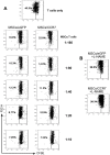
(A) CFSE labeled CD3+T cells were cultured with MSCs at different ratios in the presence of PMA plus ION for 48 hours. T cell proliferation as indicated by the reduction in CFSE intensity was analyzed by FCM. (B) L-NMMA was added at the beginning of co-culture (MSCs:T cells = 1∶10). The cells were subjected to FCM for T-cell proliferation detection. Numbers adjacent indicate percentage of cells in the gate. Data are representative of three independent experiments.
Figure 4
Figure 4. MSCs/CCR7 relocate within the T cell-rich zones of SLOs.
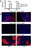
(A)MSCs/eGFP or MSCs/CCR7 were intravenously injected into GvHD mice. Five days later, the proportion of eGFP+ cells in spleen (SP) and lymphoid nodes (LN) were examined by FCM. *, p<.005. (B) Cryosection slices of SP and LN of the recipients were immunofluorescent stained with CD3 antibody (red, upper panel), CD11c antibody (red, middle panel) or B220 antibody (red, lower panel), then counterstained with DAPI (blue). Bar = 100 µm. Data were representative of three independent experiments.
Figure 5
Figure 5. MSCs/CCR7 infusion remarkably inhibits the development of GvHD.
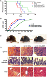
(A) Survival curve of each group of mice (n = 12). (B) The degree of systemic GvHD scores of mice (n = 12). (C) The representative clinical experience of the three groups of mice.(D) The representative pathologic changes at day 14. Original magnification, ×200. Data were representative of three independent experiments.
Figure 6
Figure 6. MSCs/CCR7 co-infusion makes the T cells more naïve like and thus coerce them stay in SLOs from trafficking to the target organs.
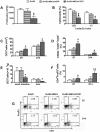
(A) Splenic T cells were collected for proliferation capacity detection by 3H-thymidine uptake. (B) The cytotoxity of the splenic T cells of the three groups of mice. (C) The absolute numbers of the CD3+T cells in spleens of the three groups of mice, which were got by multiplying the nucleocyte numbers with the CD3+ cell percentage examined by FCM; (D) The proportion of the CD62L+CCR7+ cells in CD3+ T lymphocytes; (E) The absolute numbers of the CD3+ cells in the small intestines and livers of the three groups of mice. (F) The proportion of CD4+FoxP3+ Tregs in spleens of the three groups of mice; (G)The respective FCM results in (F) Data shown are mean±S.D. of a representative of 3 independent experiments.*P<0.05 when compared with the GvHD group of mice.

References

    1. Billingham RE (1966–1967) The biology of graft-versus-host reactions. Harvey Lect 62:21–78. - PubMed
    1. Kim YM, Sachs T, Asavaroengchai W, Bronson R, Megan Sykes M, et al. (2003) Graft-versus-host disease can be separated from graft-versus-lymphoma effects by control of lymphocyte trafficking with FTY720. J Clin Invest 111:659–669. - PMC - PubMed
    1. Taylor PA, Ehrhardt MJ, Lees CJ, Tolar J, Weigel BJ, et al. (2007) Insights into the mechanism of FTY720 and compatibility with regulatory T cells for the inhibition ofgraft-versus-host disease (GVHD). Blood 110:3480–8. - PMC - PubMed
    1. Kataoka H, Ohtsuki M, Shimano K, Mochizuki S, Oshita K, et al. (2005) Immunosuppressive activity of FTY720, sphingosine 1-phosphate receptor agonist: II. Effect of FTY720 and FTY720-phosphate on host-versus-graft and graft-versus-host reaction in mice. Transplant Proc 37:107–109. - PubMed
    1. Sackstein R (2006) A revision of Billingham’s tenets: The central role of lymphocyte migration in acute graft-versus-host disease. Biol Blood Marrow Transplant 12:2–8. - PubMed

Publication types

LinkOut - more resources