Review
. 2015 Feb;16(2):95-109.
doi: 10.1038/nrm3918. Epub 2014 Dec 30.

In the right place at the right time: visualizing and understanding mRNA localization

Affiliations
Review

In the right place at the right time: visualizing and understanding mRNA localization

Adina R Buxbaum et al. Nat Rev Mol Cell Biol. 2015 Feb.

Erratum in

  • Nat Rev Mol Cell Biol. 2015 Aug;16(8):513

Abstract

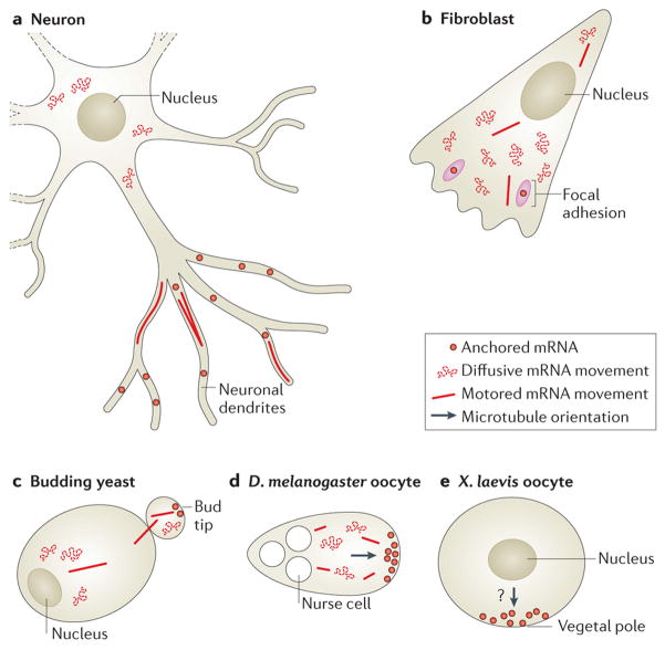
The spatial regulation of protein translation is an efficient way to create functional and structural asymmetries in cells. Recent research has furthered our understanding of how individual cells spatially organize protein synthesis, by applying innovative technology to characterize the relationship between mRNAs and their regulatory proteins, single-mRNA trafficking dynamics, physiological effects of abrogating mRNA localization in vivo and for endogenous mRNA labelling. The implementation of new imaging technologies has yielded valuable information on mRNA localization, for example, by observing single molecules in tissues. The emerging movements and localization patterns of mRNAs in morphologically distinct unicellular organisms and in neurons have illuminated shared and specialized mechanisms of mRNA localization, and this information is complemented by transgenic and biochemical techniques that reveal the biological consequences of mRNA mislocalization.


Conflict of interest statement

Competing interests statement

The authors declare no competing interests.

Figures

Figure 1
Figure 1. Visualizing and understanding mRNA localization in different model systems
Aa | mRNA localization in oocytes and embryos can be essential for future patterning and development. In Drosophila melanogaster oocytes, cytoplasmic streaming from the nurse cells at the anterior drives nanos mRNA to the posterior, where it is anchored and translated, whereas mRNAs not present at the posterior are repressed to prevent translation (see Box 2 for a discussion on another mechanism contributing to the localized translation of nanos mRNA).Ab | Single-molecule fluorescence in situ hybridization (smFISH) of nanos mRNA localization in a 1-hour-old D. melanogaster embryo is shown. Ba | mRNA localization has an important role in cell fate determination during cell division. For example, in Saccharomyces cerevisiae, ASH1 mRNA, which encodes a transcription inhibitor, is transported to the bud tip by the locasome, a protein complex consisting of several RNA-binding proteins (RBPs) and a myosin motor. Local translation at the bud tip inhibits the transcription of the homothallic switching (HO) endonuclease, which is required for mating type switch, thus preventing mating type switching in the daughter cell. Bb | smFISH of ASH1 mRNA in S. cerevisiae is shown. Ca | mRNA localization and local translation in fibroblasts have been shown to be important for proper cell migration and motility. For example, β-actin mRNA localization to the cell edge is correlated with cell polarization, and β-actin mRNA localization to focal adhesions is crucial for proper cell migration. Cb | smFISH of β-actin mRNA in a cultured mouse embryonic fibroblast is shown. Cc | Enlarged image of dashed box in part Cb is shown. Da | In Escherichia coli, the bicistronic bglF–bglG transcript localizes to the plasma membrane in a translation-independent manner. This leads to the localization of BglF and BglG to the membrane. If bglG is transcribed as a monocistronic mRNA, it will localize to the poles (Fig. 4B) where, under certain conditions, the BglG protein will also be localized. Db | Image shows membrane-localized MS2–GFP-tagged endogenous bglF mRNA in E. coli. Ea | In neurons, mRNA localization to synapses is thought to be crucial for synapse development and plasticity. mRNAs, such as β-actin, are transported in dendrites, and synaptic activity is proposed to cause mRNAs to localize at stimulated synapses. The local translation of β-actin is proposed to cause morphological and functional alterations of synapses. Eb | smFISH of β-actin mRNA in a cultured mouse hippocampal neuron is shown. Ec | Enlarged image of dashed box in part Eb is shown. Each side of the dashed boxes of parts Ab, Cc and Ecrepresents 20 μm. Image in part Ab courtesy of T. Trcek and R. Lehmann, New York University School of Medicine, USA. Image in part Bb courtesy of T. Trcek. Image in part Db courtesy of O. Amster-Choder, The Hebrew University of Jerusalem, Israel.
Figure 2
Figure 2. Traditional and novel uses of MS2-like systems to investigate mRNA biology
a | Localization of single mRNA molecules can be studied by tagging with fluorescent proteins. The fusion of a stem–loop-binding protein, for example, the phage MS2 coat protein (MCP), to a fluorescent protein such as GFP allows single-molecule mRNA imaging, . b | In dual-colour labelling, two mRNAs (top) or two different parts of the same mRNA (usually the 3′ and the 5′; bottom) aretagged by different stem–loop–RNA-binding protein (RBP)–fluorescent protein systems, thereby allowing the imaging of two different mRNAs in the same cell or the analysis of RNA dynamics, such as transcription, nuclear export and degradation. c | A ‘background-free’ system is shown. To reduce background fluorescence, two different stem–loop species (for example, those of the MS2 and PP7 phage systems) bound to their respective RBPs — MCP and PP7 coat protein (PCP) — are used. Each RBP is fused to one half of a split yellow fluorescent protein (YFP), which by itself does not fluoresce. Only when both MCP and PCP are bound to the mRNA are the two halves close enough to become a functional YFP. d | The tethering of an mRNA to a specific cellular location or structure (for example, focal adhesions) is carried out by fusing MCP to a protein (in this case, vinculin) with specific subcellular localization that anchors the mRNA to a specific cellular compartment, body or organelle. e | In RNA affinity purification, an RBP such as MCP is fused to a unique epitope — for example, streptavidin-binding protein (SBP) — which mediates the affinity purification of the RNA (along with mRNA – protein (mRNP) complexes that might bind to it) using streptavidin and biotin beads. f | A specific protein can be tethered to an mRNA through RBPs. The protein in question, which is thought to affect the mRNA or its associated proteins (for example, the transporter She3), is fused to MCP, which tethers it to the mRNA and allows the analysis of this direct interaction. g | Simultaneous localization of the mRNA and its protein product can also be studied. By fusing the gene to the mCherry (a red fluorescent protein) open reading frame (ORF) and cloning MS2-binding sites (MBSs) into its 3′ untranslated region (UTR), the mRNA can be visualized by MCP – GFP binding and the protein by mCherry fluorescence. See Supplementary information S2 (table) for more information.
Figure 3
Figure 3. Cellular determinants of motored mRNA transport
Owing to the bidirectional orientation of microtubules in most cell types and to the unidirectional movement of each molecular motor along them, the directional movement of mRNA in cells may seem disorganized when observed. Nevertheless, several cellular mechanisms of biased motored mRNA transport have recently been identified. a | To increase the processivity of directed mRNA transport, some mRNAs bind to multiple motors. For example, each ASH1 mRNA molecule has four localization elements, which mediate the binding of four She3 RNA-binding proteins (RBPs) and, in turn, the binding of four myosins. b |Local biases in the orientation of microtubules have been shown to cause a bias in mRNA transport, allowing mRNA localization, , . c | In the case of mixed-orientation microtubules, mRNAs bound to multiple motors may experience a ‘tug of war’ and will be transported in the direction of the strongest combined motor force. d | Alternatively, mRNAs bound by different types of molecular motors, which move in opposite directions, may also undergo a tug of war and will be transported in the direction of the stronger force exerted. e | Microtubule-associated proteins (MAP) have been shown to alter the dissociation rates of motors from microtubules and to cause a motor to change direction when moving along a microtubule, presumably by behaving as an obstacle, . f | The binding of cargoes to motors has been shown to alter their binding and motility on microtubules, in addition to increasing their processivity along microtubules, . mRNP, mRNA – protein.
Figure 4
Figure 4. mRNA localization in unicellular organisms
A | In budding yeast, the ASH1 and SRO7 mRNAs are transported to the bud tip by the locasome (which comprises Myo4, She3 and She2), Scp160 and Puf6 (part Aa). Following pheromone chemotaxis, SRO7 mRNA is transported to the shmoo tip by Scp160 but not by the locasome (part Ab). mRNAs encoding membrane or secreted proteins (such as USE1 and SUC2) are localized to the endoplasmic reticulum (ER), in a Puf2- and She2-dependent manner (part Ac), whereas the OXA1 and ATP2 mRNAs, which encode mitochondrial proteins, are targeted to mitochondria or to the mitochondrion–ER interface in a Puf3-dependent manner (part Ad). Some mRNAs encoding peroxisomal proteins (for example, PEX1, PEX5 and PEX14) are localized to peroxisomes in a Puf5-dependent manner (part Ae). The ABP140 mRNA, which encodes AdoMet-dependent tRNA methyltransferase, is transported to the far pole of the mother cell by direct binding of the amino terminus of its nascent protein product, Abp140, to actin filaments. The retrograde movement of actin drives ABP140 mRNA to the far pole in a motor-independent manner (part Af). B | In bacteria, the Escherichia coli lacY and bglG–bglF mRNAs, which encode transmembrane proteins, localize to the plasma membrane (part Ba). bglG transcribed as a monocistronic mRNA localizes to the cell poles (part Bb), whereas bglB transcribed alone is cytoplasmic (part Bc). In Bacillus subtilis, the comE transcript, which is an operon that encodes factors for horizontal gene transfer, is localized to the nascent septum that separates daughter cells, and to cell poles (part Bd).
Figure 5
Figure 5. Different types of mRNA movements depend on subcellular location and on cell type
a | Different types of mRNA movements can be observed in neurons, including diffusive movement; active, motored transport; and stalling or anchoring of mRNAs. Whereas around the nucleus, mRNAs encoding β-actin seem to move in a diffusive manner, in dendrites β-actin and ARC mRNAs seem to be largely stalled or corralled, and 10% of dendritic mRNAs are seen to undergo active transport on microtubules, , , . b | In fibroblasts, most β-actin mRNAs are diffusing. A small percentage is transported along microtubules, and some mRNAs dwell near focal adhesions, . c | In budding yeast, ASH1 mRNAs are mostly diffusive. The localization of the ASH1 mRNA is accomplished through myosin-mediated transport to the bud tip, where the mRNA is anchored, , .d | oskar mRNAs in Drosophila melanogaster oocytes move around mostly in a diffusive manner. A small percentage can be seen moving along the cytoskeleton for brief lengths. At the posterior, oskar mRNAs are anchored. e | vg1 mRNAs in Xenopus laevis oocytes localize to the vegetal cortex owing to a bias in the placement of the plus ends of microtubules. This localization depends on two forms of kinesin, although the precise dynamics of vg1 mRNA movement are unclear.

Comment in

References

    1. Weatheritt RJ, Gibson TJ, Babu MM. Asymmetric mRNA localization contributes to fidelity and sensitivity of spatially localized systems. Nature Struct Mol Biol. 2014;21:833–839. - PMC - PubMed
    1. Jeffery WR, Tomlinson CR, Brodeur RD. Localization of actin messenger RNA during early ascidian development. Dev Biol. 1983;99:408–417. - PubMed
    1. Lawrence JB, Singer RH. Intracellular localization of messenger RNAs for cytoskeletal proteins. Cell. 1986;45:407–415. - PubMed
    1. Melton DA. Translocation of a localized maternal mRNA to the vegetal pole of Xenopusoocytes. Nature. 1987;328:80–82. - PubMed
    1. Berleth T, et al. The role of localization of bicoid RNA in organizing the anterior pattern of the Drosophila embryo. EMBO J. 1988;7:1749–1756. - PMC - PubMed

Publication types