. 2015 Mar;352(3):509-18.
doi: 10.1124/jpet.114.221929. Epub 2014 Dec 30.

Cytokine-induced S-nitrosylation of soluble guanylyl cyclase and expression of phosphodiesterase 1A contribute to dysfunction of longitudinal smooth muscle relaxation

Affiliations

Cytokine-induced S-nitrosylation of soluble guanylyl cyclase and expression of phosphodiesterase 1A contribute to dysfunction of longitudinal smooth muscle relaxation

Senthilkumar Rajagopal et al. J Pharmacol Exp Ther. 2015 Mar.

Abstract

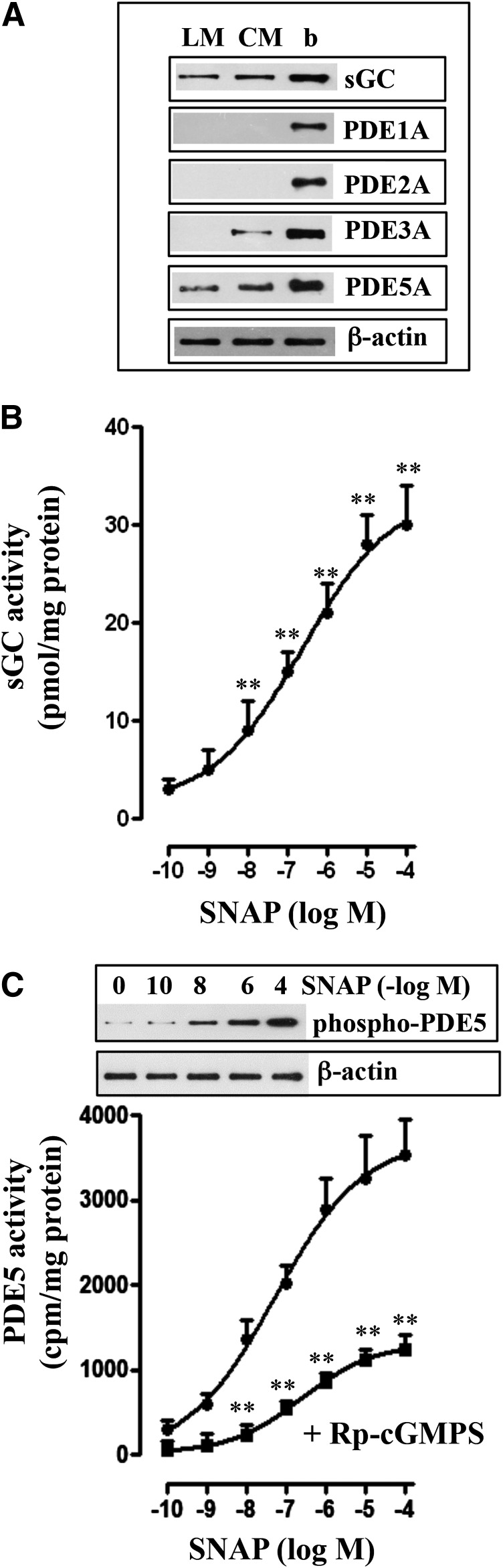
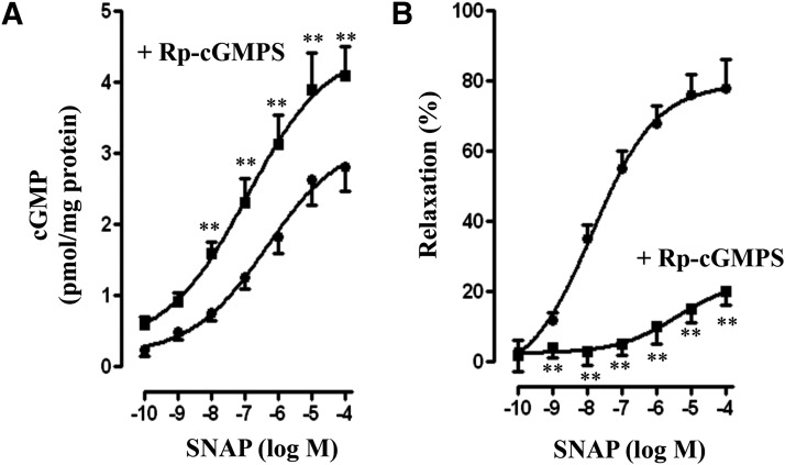
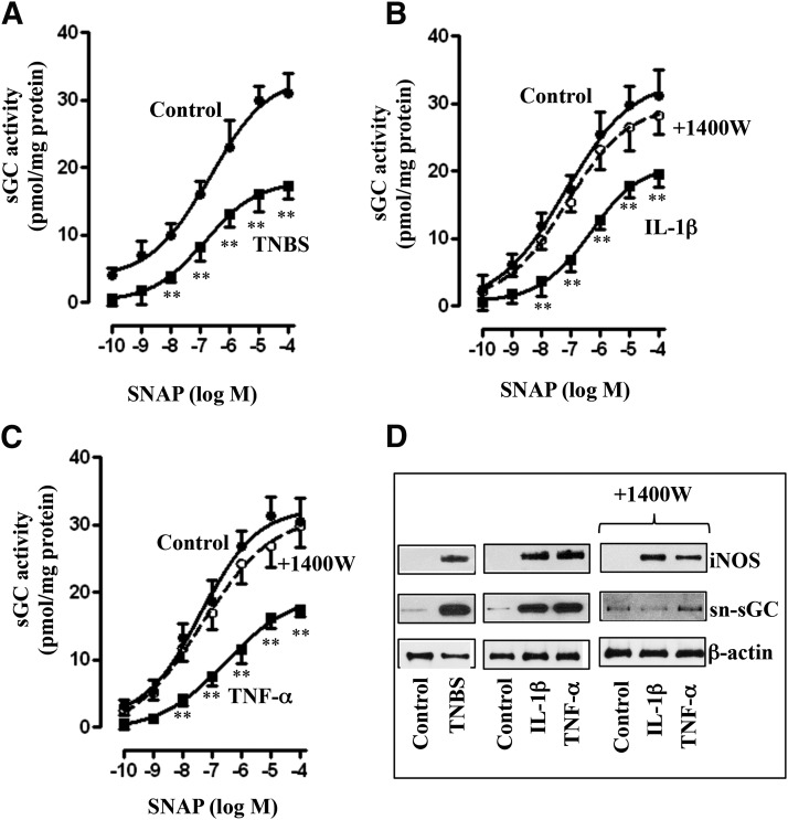
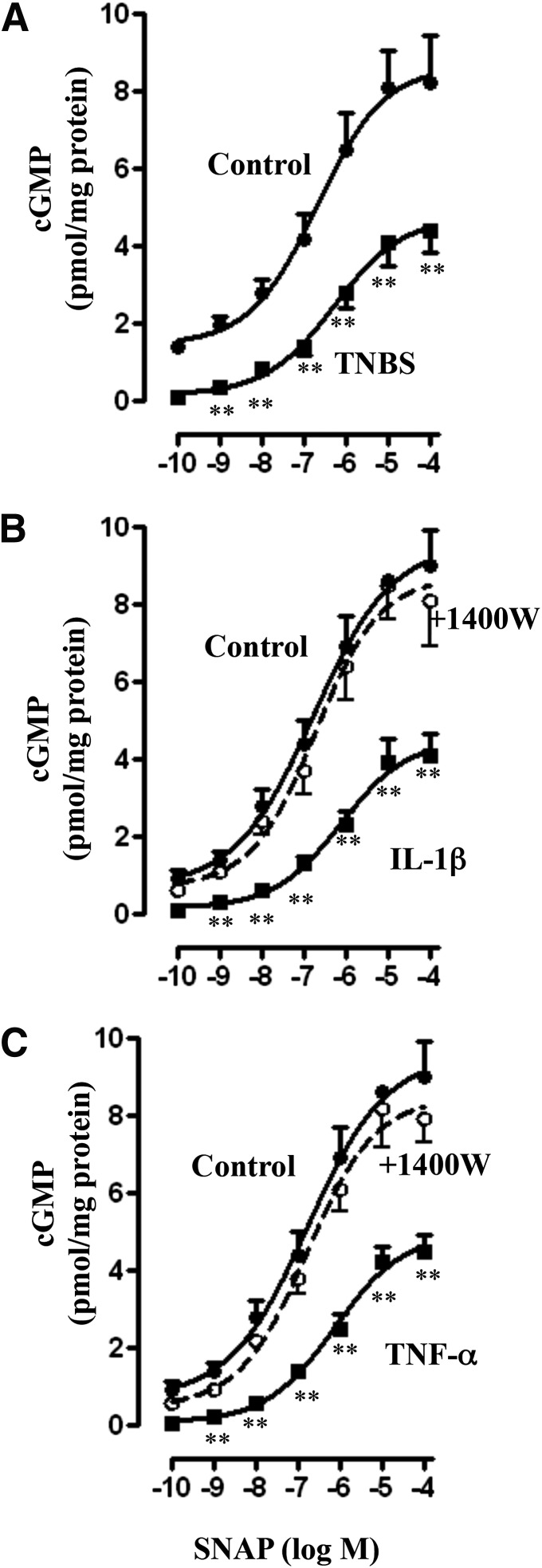
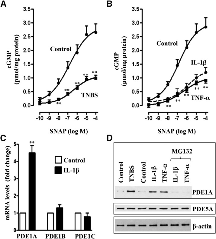
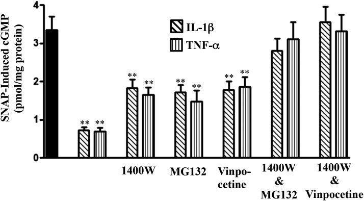
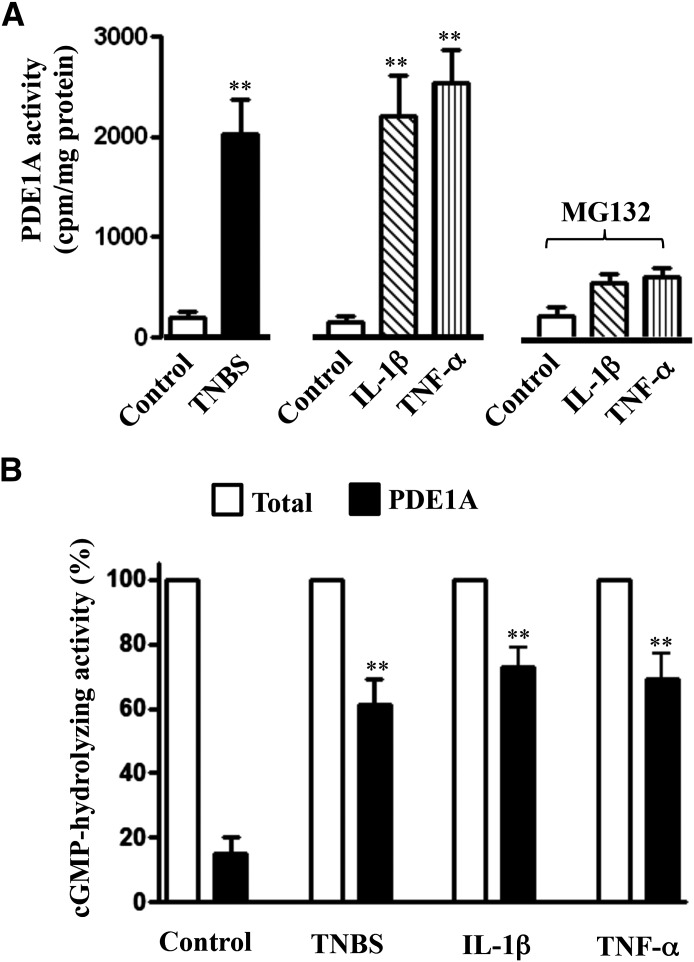
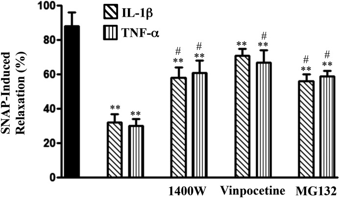
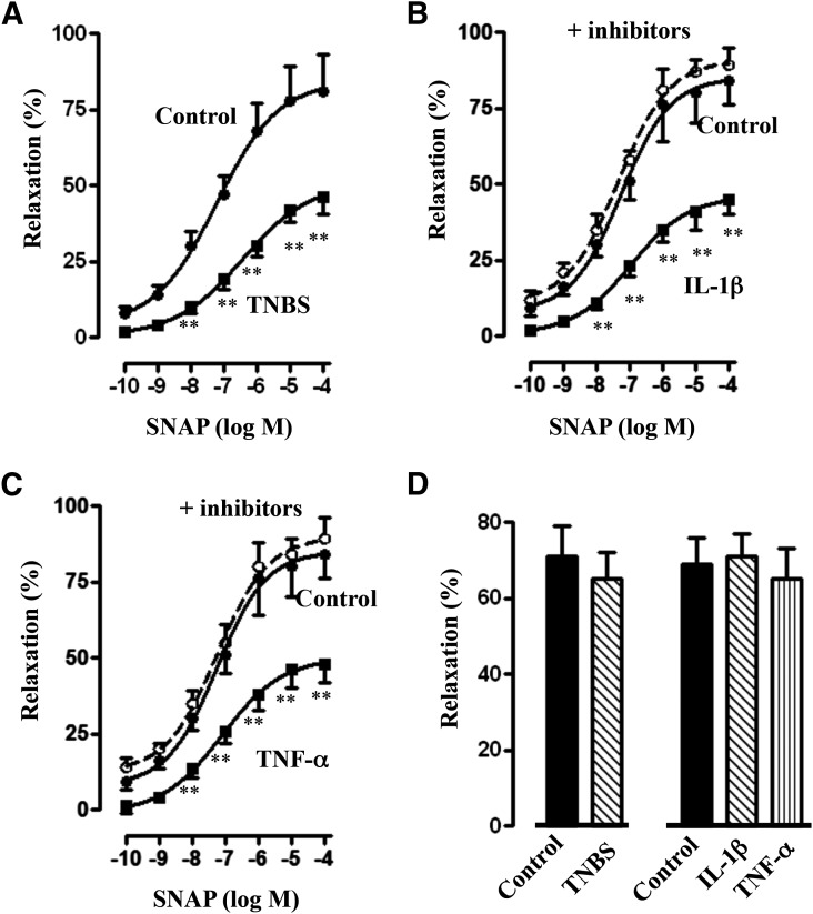
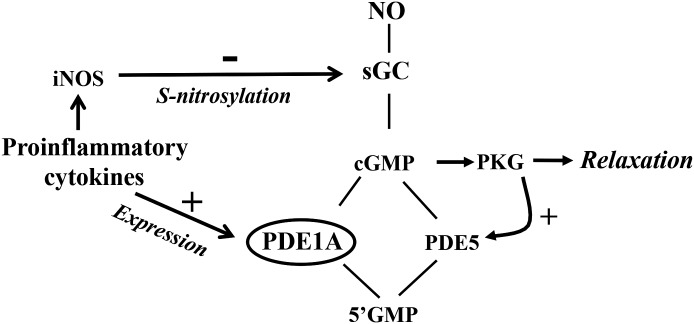
The effect of proinflammatory cytokines on the expression and activity of soluble guanylyl cyclase (sGC) and cGMP-phosphodiesterases (PDEs) was determined in intestinal longitudinal smooth muscle. In control muscle cells, cGMP levels are regulated via activation of sGC and PDE5; the activity of the latter is regulated via feedback phosphorylation by cGMP-dependent protein kinase. In muscle cells isolated from muscle strips cultured with interleukin-1β (IL-1β) or tumor necrosis factor α (TNF-α) or obtained from the colon of TNBS (2,4,6-trinitrobenzene sulfonic acid)-treated mice, expression of inducible nitric oxide synthase (iNOS) was induced and sGC was S-nitrosylated, resulting in attenuation of nitric oxide (NO)-induced sGC activity and cGMP formation. The effect of cytokines on sGC S-nitrosylation and activity was blocked by the iNOS inhibitor 1400W [N-([3-(aminomethyl)phenyl]methyl)ethanimidamide dihydrochloride]. The effect of cytokines on cGMP levels measured in the absence of IBMX (3-isobutyl-1-methylxanthine), however, was partly reversed by 1400W or PDE1 inhibitor vinpocetine and completely reversed by a combination of 1400W and vinpocetine. Expression of PDE1A was induced and was accompanied by an increase in PDE1A activity in muscle cells isolated from muscle strips cultured with IL-1β or TNF-α or obtained from the colon of TNBS-treated mice; the effect of cytokines on PDE1 expression and activity was blocked by MG132 (benzyl N-[(2S)-4-methyl-1-[[(2S)-4-methyl-1-[[(2S)-4-methyl-1-oxopentan-2-yl]amino]-1-oxopentan-2-yl]amino]-1-oxopentan-2-yl]carbamate), an inhibitor of nuclear factor κB activity. NO-induced muscle relaxation was inhibited in longitudinal muscle cells isolated from muscle strips cultured with IL-1β or TNF-α or obtained from the colon of TNBS-treated mice, and this inhibition was completely reversed by the combination of both 1400W and vinpocetine. Inhibition of smooth muscle relaxation during inflammation reflects the combined effects of decreased sGC activity via S-nitrosylation and increased cGMP hydrolysis via PDE1 expression.


Figures

Fig. 1.
Fig. 1.
Expression of sGC and cGMP-hydrolyzing PDEs in circular and longitudinal smooth muscle and SNAP-induced stimulation of sGC and PDE5 activity. (A) Expression of sGC β1 subunit and PDE isoforms were analyzed in freshly prepared dispersed circular (CM) and longitudinal (LM) smooth muscle of colon and brain (b). Lysates containing equal amounts of total proteins were separated with SDS-PAGE, and expression of sGC β1 subunit, PDE1A, PDE2A, PDE3A, and PDE5A was analyzed using selective antibody. Membranes were reblotted to measure β-actin. Protein bands were visualized with enhanced chemiluminescence. (B) Longitudinal muscle cells were treated with different concentrations of SNAP for 5 minutes. sGC activity was measured as described in Materials and Methods. Results are expressed as pmol cGMP/mg protein above basal levels (2.82 pmol/mg protein). Values are the means ± S.E.M. of five experiments. **P < 0.01, significant increase in sGC activity. (C) Longitudinal muscle cells were treated with different concentrations of SNAP in the presence or absence of PKG inhibitor, Rp-cGMPS (10 µM), for 5 minutes, and PDE5 activity was measured in PDE5A immunoprecipitates as described in Materials and Methods and expressed as cpm/mg protein above basal values (295 ± 42 cpm/mg protein). Values are the means ± S.E.M. of five experiments. **P < 0.01, significant inhibition of SNAP-stimulated PDE5 activity by Rp-cGMPS. Inset: Representative immunoblot of four different experiments. Longitudinal muscle cells were treated with different concentrations of SNAP, and PDE5A immunoprecipitates were separated on SDS-PAGE and analyzed with phospho-specific (Ser92) antibody in the Western blot.
Fig. 2.
Fig. 2.
SNAP-induced cGMP generation and muscle relaxation. (A) Longitudinal muscle cells were treated with different concentrations of SNAP in the presence or absence of Rp-cGMPS (10 µM) for 5 minutes. cGMP was measured in the presence of 100 µM IBMX by radioimmunoassay and expressed as pmol/mg protein above basal levels (0.22 ± 0.04 pmol/mg protein). Values are the means ± S.E.M. of four experiments. **P < 0.01, significant increase in SNAP-induced cGMP generation by Rp-cGMPS. (B) Longitudinal muscle cells isolated from colon were treated with different concentrations of SNAP in the presence or absence of Rp-cGMPS (10 µM) for 5 minutes followed by carbachol for 30 seconds to measure initial Ca2+-dependent contraction. Smooth muscle cell contraction was measured by scanning micrometry, and relaxation was expressed as the percent inhibition of carbachol-induced contraction (basal cell length: 95 ± 4 µm; carbachol-induced contraction: 31 ± 3% decrease in cell length). Values are the means ± S.E.M. of five to six experiments. **P < 0.01 significant inhibition in SNAP-induced relaxation by Rp-cGMPS.
Fig. 3.
Fig. 3.
Suppression of SNAP-induced sGC activity via iNOS-mediated nitrosylation of sGC. Longitudinal muscle cells isolated from colons of control and TNBS-treated mice (A) or from muscle strips cultured in the presence of IL-1β (10 ng/ml) (B) or TNF-α (1 nM) (C) for 48 hours were treated with different concentrations of SNAP, and sGC activity was measured as described in Materials and Methods. In some experiments, muscle strips were cultured in the presence of IL-1β (10 ng/ml) (B) or TNF-α (1 nM) (C) plus iNOS inhibitor 1400W (10 µM) for 48 hours. Basal sGC activity was not significantly different between control and TNBS-treated mice (2.61 ± 0.32 versus 2.54 ± 0.36 pmol/mg protein) or between control and cytokine-treated muscle strips (2.48 ± 0.36 versus 2.62 ± 0.36 pmol/mg protein). Values are the means ± S.E.M. of five experiments. **P < 0.01, significant inhibition of SNAP-induced sGC activity. (D) Representative immunoblot of four to five different experiments. Longitudinal muscle cells isolated from colons of control and TNBS-treated mice or from muscle strips cultured in the presence of IL-1β (10 ng/ml) or TNF-α (1 nM) for 48 hours. In some experiments, muscle strips were cultured with IL-1β (10 ng/ml) or TNF-α (1 nM) in the presence of iNOS inhibitor 1400W (10 µM) for 48 hours. Lysates were used to measure iNOS expression and S-nitrosylation of sGC (sn-sGC) as described in Materials and Methods.
Fig. 4.
Fig. 4.
Suppression of SNAP-induced cGMP levels via iNOS. Longitudinal muscle cells isolated from colons of control and TNBS-treated mice (A) or from muscle strips cultured in the presence of IL-1β (10 ng/ml) (B) or TNF-α (1 nM) (C) for 48 hours were treated with different concentrations of SNAP. In some experiments, muscle strips were cultured with IL-1β (10 ng/ml) or TNF-α (1 nM) in the presence of iNOS inhibitor 1400W (10 µM) for 48 hours. cGMP levels were measured in the presence of 100 µM IBMX as described in Materials and Methods. Basal cGMP levels were not significantly different between control and TNBS-treated mice (0.24 ± 0.05 versus 0.21 ± 0.04 pmol/mg protein) or between control and cytokine-treated muscle strips (0.22 ± 0.04 versus 0.26 ± 0.05 pmol/mg protein). Values are the means ± S.E.M. of five experiments. **P < 0.01, significant inhibition in SNAP-induced cGMP formation.
Fig. 5.
Fig. 5.
Upregulation of PDE1A expression via NF-κB and suppression of SNAP-induced cGMP levels. Longitudinal muscle cells isolated from colons of control and TNBS-treated mice (A) or from muscle strips cultured in the presence of IL-1β (10 ng/ml) or TNF-α (1 nM) (B) for 48 hours were treated with different concentrations of SNAP. Cyclic GMP levels were measured in the absence of IBMX as described in Materials and Methods. Basal cGMP levels were not significantly different between control and TNBS-treated mice or between control and cytokine-treated muscle strips. Values are the means ± S.E.M. of five experiments. **P < 0.01, significant inhibition of SNAP-induced cGMP formation. (C) Longitudinal muscle cells in culture were treated with IL-1β for 48 hours, and expression of PDE1 isoforms was measured by real-time PCR as described in Materials and Methods. Values are the means ± S.E.M. of five experiments. **P < 0.01, significant increase in PDE1A expression. (D) Representative immunoblot of four to five different experiments. Longitudinal muscle cells were isolated from colons of control and TNBS-treated mice or from muscle strips cultured in the presence of IL-1β (10 ng/ml) or TNF-α (1 nM) for 48 hours. In some experiments, muscle strips were cultured with IL-1β (10 ng/ml) or TNF-α (1 nM) in the presence of NF-κB inhibitor MG132 (10 µM) for 48 hours. Expression of PDE1A and PDE5 was analyzed by Western blot.
Fig. 6.
Fig. 6.
Suppression of SNAP-induced cGMP formation by proinflammatory cytokines via iNOS- and NF-κB–mediated PDE1A activity. Longitudinal muscle cells were isolated from muscle strips cultured in the presence of IL-1β (10 ng/ml) or TNF-α (1 nM) for 48 hours. In some experiments, muscle strips were cultured with IL-1β (10 ng/ml) or TNF-α (1 nM) in the presence of NF-κB inhibitor MG132 (10 µM), iNOS inhibitor 1400W (10 µM), PDE1 inhibitor vinpocetine (50 µM), or 1400W in combination with MG132 or vinpocetine for 48 hours. cGMP levels in response to SNAP (10 µM) were measured in the absence of IBMX by radioimmunoassay. Values are the means ± S.E.M. of five experiments. **P < 0.01, significant inhibition compared with control SNAP-induced cGMP formation (solid bar).
Fig. 7.
Fig. 7.
Increase in PDE1A activity by inflammation. (A) Longitudinal muscle cells isolated from colons of control and TNBS-treated mice or from muscle strips cultured in the presence of IL-1β (10 ng/ml) or TNF-α (1 nM) for 48 hours were used to measure PDE1A activity as described in Materials and Methods. In some experiments, muscle strips were cultured with IL-1β (10 ng/ml) or TNF-α (1 nM) in the presence of NF-κB inhibitor MG132 (10 µM) for 48 hours. Values are the means ± S.E.M. of five experiments. **P < 0.01, significant increase in PDE1A activity compared with control. (B) Longitudinal muscle cells isolated from colons of control and TNBS-treated mice or from muscle strips cultured in the presence of IL-1β (10 ng/ml) or TNF-α (1 nM) for 48 hours were used to measure cGMP-hydrolyzing activity in the presence or absence of PDE1 inhibitor vinpocetine (50 µM). Total cGMP-hydrolyzing activity reflects the activity in the absence of vinpocetine. PDE1A activity was calculated as the percentage of total cGMP-hydrolyzing activity that was inhibited by vinpocetine. Values are the means ± S.E.M. of five experiments. **P < 0.01, significant increase in PDE1A activity compared with control.
Fig. 8.
Fig. 8.
Suppression of SNAP-induced relaxation by proinflammatory cytokines and reversal of inhibition by blockade of iNOS, NF-κB, or PDE1 activity. Longitudinal muscle cells were isolated from muscle strips cultured in the presence of IL-1β (10 ng/ml) or TNF-α (1 nM) for 48 hours. In some experiments, muscle strips were cultured with IL-1β or TNF-α in the presence of 1400W (10 µM), vinpocetine (50 µM), or MG132 (10 µM) for 48 hours. Cells were treated with 10 µM SNAP for 5 minutes followed by carbachol for 30 seconds to measure initial Ca2+-dependent contraction. Smooth muscle cell contraction was measured by scanning micrometry, and relaxation was expressed as the percent inhibition of carbachol-induced contraction. Values are the means ± S.E.M. of five to six experiments. **Significant inhibition in SNAP-induced relaxation by IL-1β or TNF-α compared with control SNAP-induced relaxation (solid bar). #P < 0.05, significant reversal of inhibition by 1400W, vinpocetine, or MG132.
Fig. 9.
Fig. 9.
Suppression of SNAP-induced relaxation by inflammation and complete reversal of inhibition by blockade of iNOS and PDE1 activity. Longitudinal muscle cells isolated from colons of control and TNBS-treated mice (A) or from muscle strips cultured in the presence of IL-1β (10 ng/ml) (B) or TNF-α (1 nM) (C) for 48 hours were treated with different concentrations of SNAP for 5 minutes and carbachol for 30 seconds to measure initial Ca2+-dependent contraction. In some experiments, muscle strips were cultured with IL-1β or TNF-α in the presence of both iNOS (1400W, 10 µM) and PDE1 (vinpocetine, 50 µM) inhibitors. Smooth muscle cell contraction was measured by scanning micrometry, and relaxation was expressed as the percent inhibition of carbachol-induced contraction. Values are the means ± S.E.M. of five to six experiments. **P < 0.01, significant inhibition of SNAP-induced relaxation. (D) Longitudinal muscle cells isolated from colons of control and TNBS-treated mice or from muscle strips cultured in the presence of IL-1β (10 ng/ml) or TNF-α (1 nM) for 48 hours were treated with PDE-resistant 8-Bromo-cGMP (10 µM) for 5 minutes and carbachol (1 µM) for 30 seconds to measure initial Ca2+-dependent contraction. Smooth muscle cell contraction was measured by scanning micrometry, and relaxation was expressed as the percent inhibition of carbachol-induced contraction. Values are the means ± S.E.M. of four experiments.
Fig. 10.
Fig. 10.
Schematic diagram demonstrating the effects of proinflammatory cytokines on pathways that regulate cGMP levels in colonic longitudinal smooth muscle cells. In longitudinal smooth muscle, cGMP levels are regulated by the activities of sGC and PDE5; the activity of the latter is stimulated via PKG-mediated phosphorylation. Proinflammatory cytokines inhibited sGC activity via iNOS-mediated S-nitrosylation, induced PDE1A expression via NF-κB, and stimulated cGMP hydrolysis, leading to suppression of cGMP formation and a decrease in muscle relaxation.

References

    1. Ahmad F, Degerman E, Manganiello VC. (2012) Cyclic nucleotide phosphodiesterase 3 signaling complexes. Horm Metab Res 44:776–785. - PubMed
    1. Akiho H, Blennerhassett P, Deng Y, Collins SM. (2002) Role of IL-4, IL-13, and STAT6 in inflammation-induced hypercontractility of murine smooth muscle cells. Am J Physiol Gastrointest Liver Physiol 282:G226–G232. - PubMed
    1. Alkahtani R, Mahavadi S, Al-Shboul O, Alsharari S, Grider JR, Murthy KS. (2013) Changes in the expression of smooth muscle contractile proteins in TNBS- and DSS-induced colitis in mice. Inflammation 36:1304–1315. - PMC - PubMed
    1. Al-Shboul O, Mahavadi S, Sriwai W, Grider JR, Murthy KS. (2013) Differential expression of multidrug resistance protein 5 and phosphodiesterase 5 and regulation of cGMP levels in phasic and tonic smooth muscle. Am J Physiol Gastrointest Liver Physiol 305:G314–G324. - PMC - PubMed
    1. Al-Shboul O, Nalli AD, Kumar DP, Zhou R, Mahavadi S, Kuemmerle JF, Grider JR, Murthy KS. (2014) Jun kinase-induced overexpression of leukemia-associated Rho GEF (LARG) mediates sustained hypercontraction of longitudinal smooth muscle in inflammation. Am J Physiol Cell Physiol 306:C1129–C1141. - PMC - PubMed

Publication types

MeSH terms

Substances

LinkOut - more resources