. 2015 Apr;29(4):1404-13.
doi: 10.1096/fj.14-261479. Epub 2014 Dec 30.

Increased Akt signaling in the mosquito fat body increases adult survivorship

Affiliations

Increased Akt signaling in the mosquito fat body increases adult survivorship

Anam J Arik et al. FASEB J. 2015 Apr.

Abstract

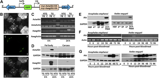
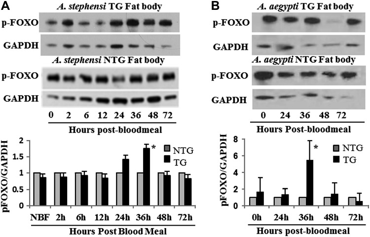
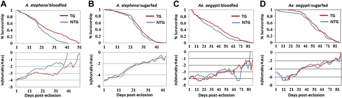
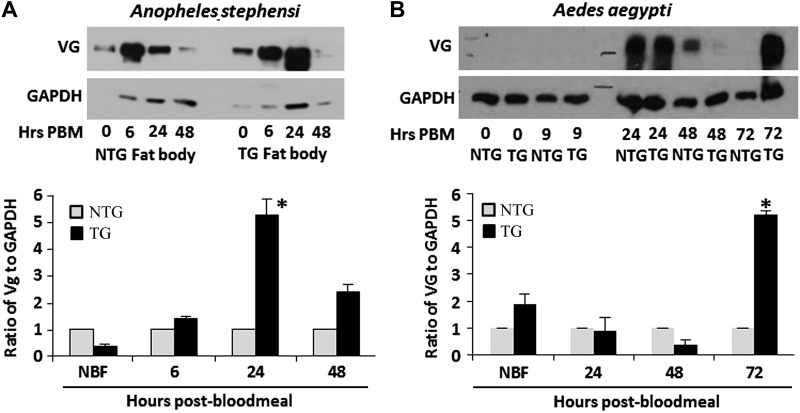
Akt signaling regulates diverse physiologies in a wide range of organisms. We examine the impact of increased Akt signaling in the fat body of 2 mosquito species, the Asian malaria mosquito Anopheles stephensi and the yellow fever mosquito Aedes aegypti. Overexpression of a myristoylated and active form of A. stephensi and Ae. aegypti Akt in the fat body of transgenic mosquitoes led to activation of the downstream signaling molecules forkhead box O (FOXO) and p70 S6 kinase in a tissue and blood meal-specific manner. In both species, increased Akt signaling in the fat body after blood feeding significantly increased adult survivorship relative to nontransgenic sibling controls. In A. stephensi, survivorship was increased by 15% to 45%, while in Ae. aegypti, it increased 14% to 47%. Transgenic mosquitoes fed only sugar, and thus not expressing active Akt, had no significant difference in survivorship relative to nontransgenic siblings. Expression of active Akt also increased expression of fat body vitellogenin, but the number of viable eggs did not differ significantly between transgenic and nontransgenic controls. This work demonstrates a novel mechanism of enhanced survivorship through increased Akt signaling in the fat bodies of multiple mosquito genera and provides new tools to unlock the molecular underpinnings of aging in eukaryotic organisms.

Keywords: Aedes aegypti; Anopheles stephensi; aging; insulin signaling; vitellogenin.


Figures

Figure 1.
Figure 1.
Generation and expression profiles of TG A. stephensi and Ae. aegypti mosquitoes with increased IIS in fat body. A) Schematic of construct engineered into TG mosquito lines. Myristoylated Akts were linked to an HA epitope to facilitate protein identification and engineered downstream of the A. gambiae or Ae. aegypti VG (VG) promoter. The EGFP fluorescent marker was linked to the synthetic 3XP3 promoter to drive expression in the eyes and nervous system. These 2 genes were flanked by the left and right arms of the piggyback (pBac) transposon. B) Example of EGFP expression in the eyes of larval TG Ae. aegypti (1) and A. stephensi (4) compared to NTG Ae. aegypti (2) and A. stephensi (3). The top panel shows the larvae under white light, the middle with a green fluorescent protein filter, and the third a merging of the top 2. C) Transcript expression in the fat body (FB) and carcass (C) of TG and NTG mosquitoes before blood feeding (0 hours) and 24 hours after blood feeding (24 hours). Actin was used as a positive control. D) Protein expression in the fat body and carcass of TG and NTG mosquitoes before blood feeding (0 hours) and 24 hours after blood feeding (24 hours). E) Expression profile of myr-AsteAkt-HA protein during mosquito development. Non-blood-fed adult females (NBF), fourth-instar larva (4th), early pupae (EP), late pupae (LP), and 24 hours after blood meal for A. stephensi (AsteAkt) and Ae. aegypti (AaegAkt). F) Postblood meal time course of transcript expression in the fat body of adult TG A. stephensi females utilizing myr-AsteAkt-specific primers (top) showed that transcript expression occurred from 12 to 36 hours after blood meal. AsteActin-specific primers were used as a positive control. Postblood meal time course of protein expression in the fat bodies of TG A. stephensi females (bottom) shows that myr-AsteAkt-HA protein is first present 24 hours after blood meal, reaches maximal expression between 36 and 48 hours after blood meal, and begins to decline by 72 hours after blood meal. GAPDH protein expression was used as a loading control. G) Postblood meal time course of transcript expression in the fat body of TG Ae. aegypti mosquitoes utilizing myr-AaegAkt-specific primers (top) showed that expression occurred at 12 and 24 hours after blood meal. AaegActin-specific primers were used as positive control. Postblood meal time course of protein expression in the fat bodies of TG A. stephensi females (bottom) shows that myr-AsteAkt-HA protein is first present 12 hours after blood meal, reaches maximal expression between 24 and 48 hours after blood meal, and begins to decline by 96 hours after blood meal. GAPDH was used as a loading control for protein analysis. All transcript and protein expression studies were replicated a minimum of 3 times.
Figure 2.
Figure 2.
Phosphorylation of the downstream IIS signaling molecule FOXO by myr-Akt. A) Fat bodies from 3 to 5 days old TG and NTG A. stephensi females were dissected to assess the phosphorylation of the downstream IIS molecule FOXO (pFOXO). Relative levels of pFOXO were measured by immunoblotting using pFOXO and GAPDH (control) antibodies (top). Shown is a representative immunoblot showing pFOXO levels throughout a reproductive cycle. In the bottom graph, the relative levels of pFOXO (determined by the ratio of pFOXO to GAPDH controls) are shown. To highlight differences between TG and NTG samples, pFOXO levels in the NTG controls were normalized to 1. B) Levels of pFOXO in TG and NTG Ae. aegypti are shown in a representative immunoblot and densitometry analysis graph as described in (A). Each experiment was replicated a minimum of 3 times. Asterisks above bar in each graph indicates a significant difference (P < 0.05) between TG and NTG relative to GAPDH control.
Figure 3.
Figure 3.
Impact of myr-Akt expression on mosquito survival. A) Representative survivorship and mortality curves of A. stephensi NTG and TG mosquitoes reared under identical conditions and provided with a blood meal 3 times a week in addition to 10% dextrose solution ad libitum (top). B) Representative survivorship and mortality curves of A. stephensi NTG and TG mosquitoes reared under the same conditions provided with only 10% dextrose solution (top). C) Representative survivorship and mortality curves of Ae. aegypti NTG and TG mosquitoes reared under identical conditions and provided with a blood meal twice weekly in addition to 10% dextrose solution ad libitum (top). D) Representative survivorship and mortality curves of Ae. aegypti NTG and TG mosquitoes reared under the same conditions provided with 10% dextrose solution (top). See Tables 1 and 2.
Figure 4.
Figure 4.
Impact of myr-Akt expression on VG synthesis. A) Representative immunoblot of VG protein levels (VG) in the abdominal body walls/fat body of A. stephensi during a reproductive cycle (top). Graph indicates relative levels of A. stephensi VG (determined by the ratio of VG to GAPDH controls). To highlight differences between TG and NTG samples, VG levels in the NTG controls were normalized to 1. B) Representative immunoblot of VG protein levels (VG) in the abdominal body walls of Ae. aegypti during a reproductive cycle (top). Immunoblots were replicated a minimum of 3 times.

References

    1. Antonova Y., Arik A., Moore W., Riehle M., Brown M. (2012) Insulin-like peptides: structure, signaling, and function. In Insect Endocrinology (Gilbert L., ed.), pp. 63–92, Elsevier, New York
    1. Toivonen J. M., Partridge L. (2009) Endocrine regulation of aging and reproduction in Drosophila. Mol. Cell. Endocrinol. 299, 39–50 - PubMed
    1. Mukhopadhyay A., Tissenbaum H. A. (2007) Reproduction and longevity: secrets revealed by C. elegans. Trends Cell Biol. 17, 65–71 - PubMed
    1. Hyun J., Hashimoto C. (2011) Physiological effects of manipulating the level of insulin-degrading enzyme in insulin-producing cells of Drosophila. Fly (Austin) 5, 53–57 - PMC - PubMed
    1. Piper M. D., Partridge L. (2007) Dietary restriction in Drosophila: delayed aging or experimental artefact? PLoS Genet. 3, e57. - PMC - PubMed

Publication types

MeSH terms

Substances

LinkOut - more resources