. 2015 Jan 13;112(2):E127-36.
doi: 10.1073/pnas.1418450112. Epub 2014 Dec 30.

β-Helical architecture of cytoskeletal bactofilin filaments revealed by solid-state NMR

Affiliations

β-Helical architecture of cytoskeletal bactofilin filaments revealed by solid-state NMR

Suresh Vasa et al. Proc Natl Acad Sci U S A. .

Abstract

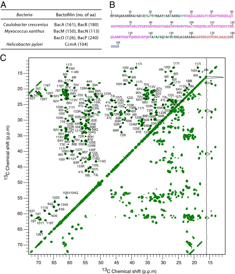
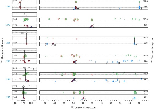
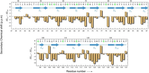
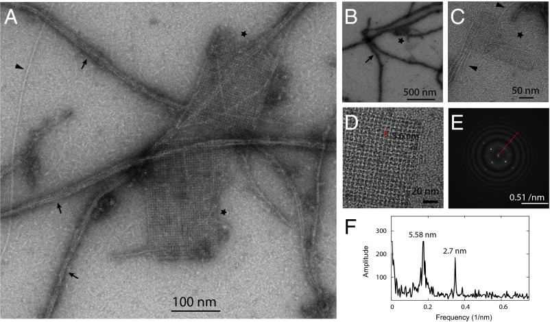
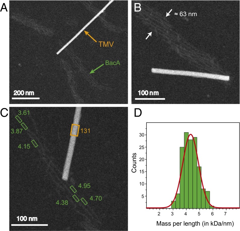
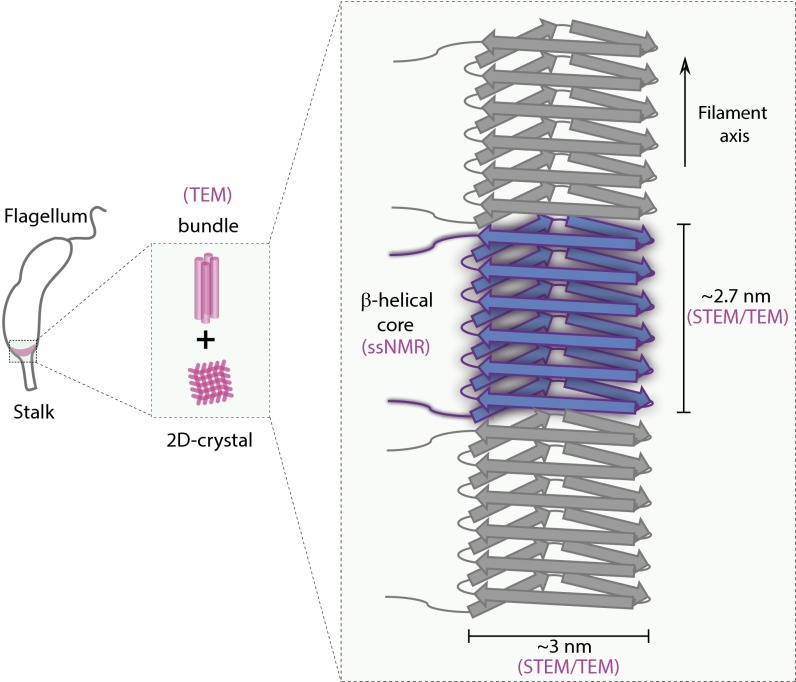
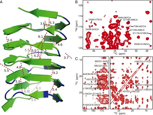
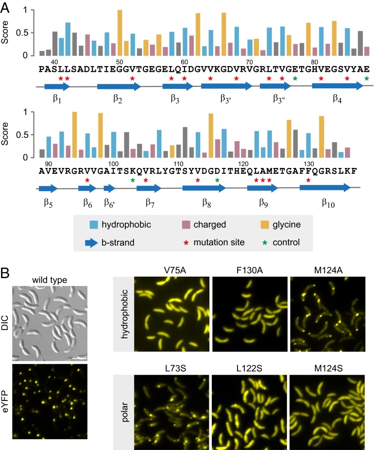
Bactofilins are a widespread class of bacterial filament-forming proteins, which serve as cytoskeletal scaffolds in various cellular pathways. They are characterized by a conserved architecture, featuring a central conserved domain (DUF583) that is flanked by variable terminal regions. Here, we present a detailed investigation of bactofilin filaments from Caulobacter crescentus by high-resolution solid-state NMR spectroscopy. De novo sequential resonance assignments were obtained for residues Ala39 to Phe137, spanning the conserved DUF583 domain. Analysis of the secondary chemical shifts shows that this core region adopts predominantly β-sheet secondary structure. Mutational studies of conserved hydrophobic residues located in the identified β-strand segments suggest that bactofilin folding and polymerization is mediated by an extensive and redundant network of hydrophobic interactions, consistent with the high intrinsic stability of bactofilin polymers. Transmission electron microscopy revealed a propensity of bactofilin to form filament bundles as well as sheet-like, 2D crystalline assemblies, which may represent the supramolecular arrangement of bactofilin in the native context. Based on the diffraction pattern of these 2D crystalline assemblies, scanning transmission electron microscopy measurements of the mass per length of BacA filaments, and the distribution of β-strand segments identified by solid-state NMR, we propose that the DUF583 domain adopts a β-helical architecture, in which 18 β-strand segments are arranged in six consecutive windings of a β-helix.

Keywords: cytoskeleton; filaments; protein structure; solid-state NMR.


Conflict of interest statement

The authors declare no conflict of interest.

Figures

Fig. 1.
Fig. 1.
High-resolution ssNMR spectra of BacA filaments. (A) Selected bacterial bactofilins. The total number of amino acids is indicated in parentheses. (B) Amino acid sequence of C. crescentus BacA, together with a synthetic linker peptide (of 17 residues, in red) and a His6-tag (in blue) attached at the C terminus. The DUF583 domain is highlighted in magenta, and prolines are shown in green. (C) Carbon–carbon 2D correlation spectrum of uniformly [13C, 15N]-labeled BacA. The carbon–carbon magnetization transfer is achieved by PDSD. A short PDSD mixing time of 20 ms was applied, optimal for intraresidue transfer. In the spectrum, a trace through Ile60 is shown to illustrate sensitivity and line width.
Fig. 2.
Fig. 2.
Sequential walk using 3D correlation spectra. Shown are strip plots of 3D NCACX (sky blue), NCOCX (light green), CANCO (black), NCACO (maroon), and NCOCA (red) spectra analyzed to perform a sequential walk along amino acids Ala128–Ala123. The connections used in the sequential walk are indicated by solid lines. The corresponding nitrogen and carbon chemical shifts are listed.
Fig. 3.
Fig. 3.
Secondary structure analysis of BacA filaments by means of secondary chemical shifts. The secondary chemical shifts (ΔδCA – ΔδCB, in ppm units) are shown as a function of the residue number for the rigid DUF583 domain. The blue arrows represent β-strands, numbered such that two continuous β-strands separated by a single glycine residue are considered as one strand. Potential kinks due to glycine residues are indicated by slashed lines. All glycine residues are highlighted in green color in the sequence.
Fig. 4.
Fig. 4.
TEM images of the uniformly [13C, 15N]-labeled BacA sample. (A) Image showing an overview of different morphologies detected by TEM. (B) BacA filament bundles (arrows). (C) 2D crystalline sheets (asterisks) and single filaments (arrowheads). (D) High-resolution image of the 2D crystalline assemblies. (E) Diffraction analysis of the 2D arrangement as obtained by Fourier transform of the image shown in D. A slice through the pattern (red dotted line) is shown in F. Individual scale bars are included in the images. For clarity, C is shown in larger format in SI Appendix, Fig. S7.
Fig. 5.
Fig. 5.
MPL measurements of BacA filaments by STEM. (A) Annular dark-field STEM image of BacA filament bundles. TMV particles are included for independent mass calibration. (B) A selected filament bundle with a width of ∼63 nm, comprising six individual filaments. (C) Filament segments used for the MPL analysis (boxes). The corresponding MPL values (in kDa/nm) are indicated. (D) MPL histogram fitted with a Gaussian function centered at 4.33 ± 0.09 kDa/nm and with an FWHH equal to 0.58 ± 0.07 kDa/nm.
Fig. 6.
Fig. 6.
Cartoon representation of the BacA filament structure. The structural parameters that were derived from the ssNMR, TEM, and STEM data collected in this work are indicated.
Fig. 7.
Fig. 7.
(A) Homology model of the DUF583 domain of BacA generated using the program Phyre2. Assigned long-range and medium-range correlations are illustrated with red dashed lines (distances in Å). For better visibility, side chains are not shown explicitly, despite the fact that some of the assigned restraints also involve side chains. Glycines are shown in blue. The cartoon representation of the β-strands follows the ssNMR secondary structure analysis (Fig. 3). (B) 2D NHHC spectrum of BacA with a 200 μs proton spin diffusion mixing time. (C) PDSD spectrum of BacA with a mixing time of 200 ms, suitable for the detection of long-range restraints. Both spectra were recorded on a 20 T (850 MHz proton Larmor frequency) wide-bore spectrometer at a magic-angle spinning rate of 11 kHz and at a temperature of 4 °C.
Fig. 8.
Fig. 8.
Mutational analysis of the BacA filament core. (A) Sequence of the BacA DUF583 domain. Numbers give the position of residues in the primary sequence of C. crescentus BacA. The bar graph indicates the conservation scores of individual residues, derived from an alignment of 150 representative bactofilin homologs, ranging from 0 (no conservation) to 1 (complete conservation). The β-strands identified in this study are shown as blue arrows. Asterisks denote residues that were targeted by site-directed mutagenesis. (B) Localization of different BacA–Venus variants in C. crescentus. Cells of strain JK5 (ΔbacAB) carrying the indicated alleles of bacA–venus under the control of the xylose-inducible Pxyl promoter were grown to late exponential phase and diluted to an OD600 of ∼0.1. After incubation for another hour, the cells were induced with 0.005% xylose for 1 h before visualization by differential interference contrast (DIC) and fluorescence microscopy. Note that wild-type BacA occasionally forms additional, nonpolar foci due to slight overexpression of the fusion protein. (Scale bar, 3 µm.)

References

    1. Bi EF, Lutkenhaus J. FtsZ ring structure associated with division in Escherichia coli. Nature. 1991;354(6349):161–164. - PubMed
    1. van den Ent F, Amos LA, Löwe J. Prokaryotic origin of the actin cytoskeleton. Nature. 2001;413(6851):39–44. - PubMed
    1. Ausmees N, Kuhn JR, Jacobs-Wagner C. The bacterial cytoskeleton: An intermediate filament-like function in cell shape. Cell. 2003;115(6):705–713. - PubMed
    1. Cabeen MT, Jacobs-Wagner C. The bacterial cytoskeleton. Annu Rev Genet. 2010;44:365–392. - PubMed
    1. Gerdes K, Howard M, Szardenings F. Pushing and pulling in prokaryotic DNA segregation. Cell. 2010;141(6):927–942. - PubMed

Publication types

MeSH terms

Substances

LinkOut - more resources