. 2015 Jan 13;112(2):566-71.
doi: 10.1073/pnas.1418580112. Epub 2014 Dec 30.

The chemistry of negotiation: rhythmic, glycan-driven acidification in a symbiotic conversation

Affiliations

The chemistry of negotiation: rhythmic, glycan-driven acidification in a symbiotic conversation

Julia A Schwartzman et al. Proc Natl Acad Sci U S A. .

Abstract

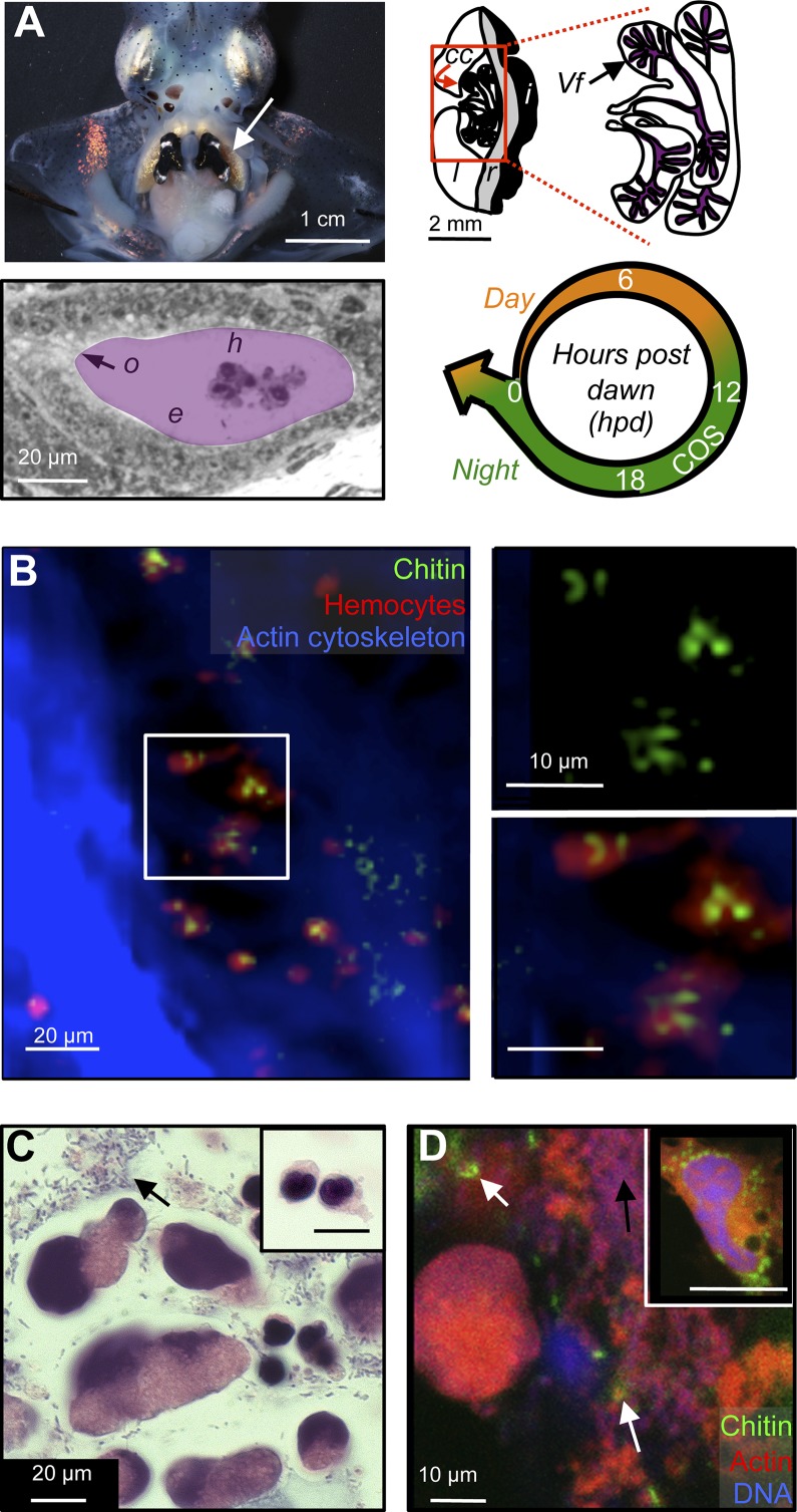
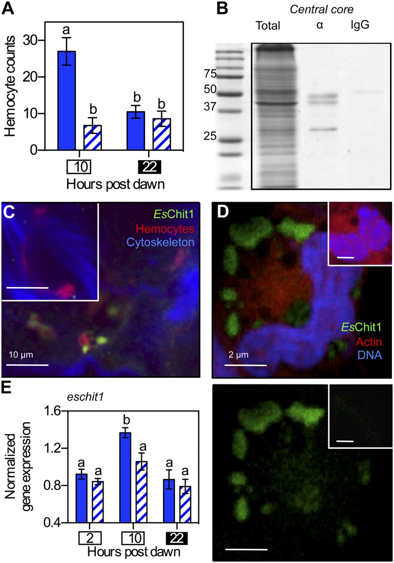
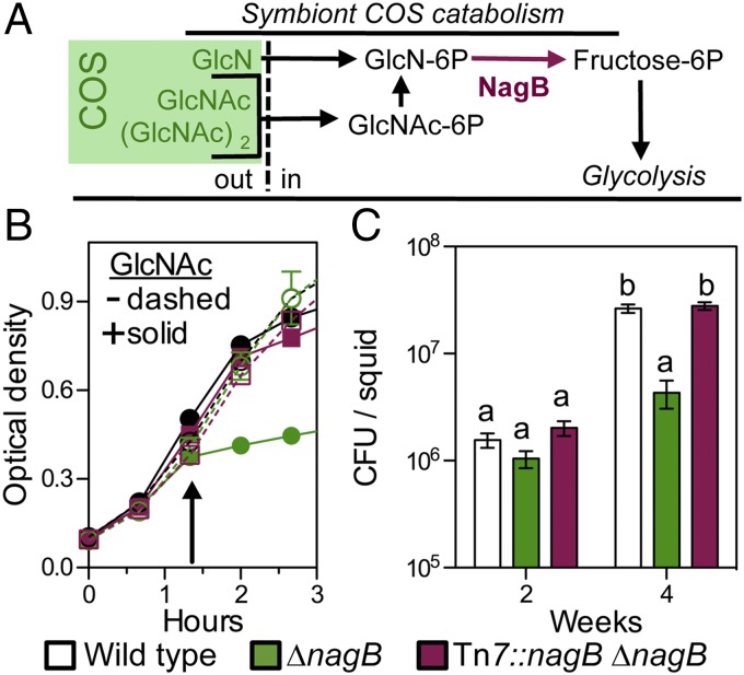
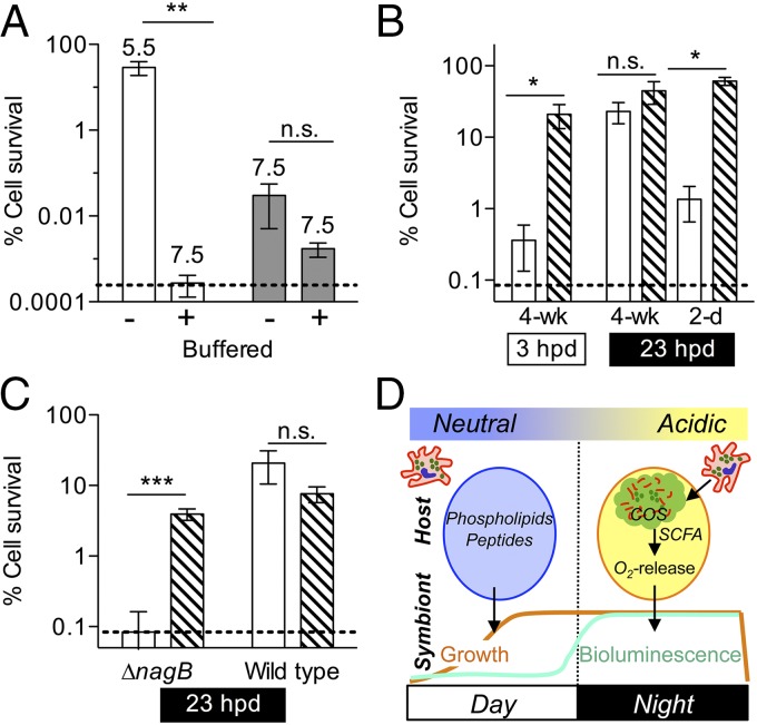
Glycans have emerged as critical determinants of immune maturation, microbial nutrition, and host health in diverse symbioses. In this study, we asked how cyclic delivery of a single host-derived glycan contributes to the dynamic stability of the mutualism between the squid Euprymna scolopes and its specific, bioluminescent symbiont, Vibrio fischeri. V. fischeri colonizes the crypts of a host organ that is used for behavioral light production. E. scolopes synthesizes the polymeric glycan chitin in macrophage-like immune cells called hemocytes. We show here that, just before dusk, hemocytes migrate from the vasculature into the symbiotic crypts, where they lyse and release particulate chitin, a behavior that is established only in the mature symbiosis. Diel transcriptional rhythms in both partners further indicate that the chitin is provided and metabolized only at night. A V. fischeri mutant defective in chitin catabolism was able to maintain a normal symbiont population level, but only until the symbiotic organ reached maturity (∼ 4 wk after colonization); this result provided a direct link between chitin utilization and symbiont persistence. Finally, catabolism of chitin by the symbionts was also specifically required for a periodic acidification of the adult crypts each night. This acidification, which increases the level of oxygen available to the symbionts, enhances their capacity to produce bioluminescence at night. We propose that other animal hosts may similarly regulate the activities of epithelium-associated microbial communities through the strategic provision of specific nutrients, whose catabolism modulates conditions like pH or anoxia in their symbionts' habitat.

Keywords: chitin; metabolism; squid–vibrio; symbiosis.


Conflict of interest statement

The authors declare no conflict of interest.

Figures

Fig. 1.
Fig. 1.
Light-organ crypts contain hemocyte-derived chitin. (A) Anatomy of the mature light organ. (Top Left) The mature, bilobed light organ is located ventrally, in the center of the mantle cavity (arrow). (Top Right) Schematic of one lobe, indicating the light-organ lens (l), reflector (r), and ink sac (i), as well as the bacteria-containing central-core tissue (cc) in the red box. Polarized epithelial cells form branched crypt spaces, in which the symbionts (Vf) reside. (Bottom Left) Symbiotic V. fischeri cells occupy extracellular crypts (purple), where they contact hemocytes (h) and the bordering epithelial cells (e); the outlet (o) to the mantle cavity allows the host to expel most of the crypt contents and symbionts every morning at dawn. (Bottom Right) The diel cycle of the mature squid–vibrio symbiosis. Throughout the animal’s life, about 95% of the symbiont population is expelled at dawn (arrow), and the remaining cells repopulate the crypts during the day (orange). At night (green), transcriptional evidence in the mature symbiosis suggests that symbionts metabolize host-derived chitin oligosaccharides (COS) (24) and produce luminescence. Numbers indicate hours post dawn (hpd). (B) Confocal micrograph, showing colocalization of chitin (fluorescent chitin-binding protein; green) and hemocytes (fluorescent DNase-I globular actin-binding protein; red) in central-core tissue (fluorescent phalloidin, a filamentous actin-binding protein; blue). Image is a 3D reconstruction of 40 1-µm confocal sections. Tissue was sampled just before nightfall (10 hpd). (Top Right and Bottom Right) Close-up of hemocytes in white box, highlighting particulate-chitin staining. (C) Differential interference contrast micrograph of crypt contents, sampled at the end of the night (22 hpd) and stained with hematoxylin (chromatin; dark blue), and eosin (proteins; pink). Two different morphologies of nucleated cells are seen in the crypt contents. Inset (same magnification) shows the single type of hemocyte morphology present in the hemolymph. Black arrow indicates extracellular material, which includes bacterial cells. (D) Detection of free particulate chitin (white arrows) and extracellular material (black arrows, here and in C) in the contents of the light-organ crypt, sampled 22 hpd. Fluorescent staining is as in B. Inset shows image of cytoplasmic particulate chitin within a hemocyte extracted from the hemolymph.
Fig. 2.
Fig. 2.
The diel migration of hemocytes to the light organ is symbiont dependent. (A) Enumeration of hemocytes present during the day (10 hpd; white box) and night (22 hpd; black box) in light-organ tissues of 2-d-old, immature symbiotic (solid bars) or aposymbiotic (hatched bars) animals. n = 15 light organs, error bars indicate SEM, and data are representative of three independent experiments. “a” and “b” indicate groups of statistically similar means, determined with two-way ANOVA with post hoc Bonferroni T-tests. (B) Western blot showing presence of EsChit1 in 25 µg of total soluble protein isolated from light-organ central cores. Total, total soluble protein; α, anti-EsChit1 antibody; IgG, Ig control. (C) Confocal micrograph localizing EsChit1 in whole adult (>4-wk-old) central cores at dusk (10 hpd). Inset shows preimmune control. (D) Localization of EsChit1 in hemocytes extracted from adult symbiotic squid. (Bottom) Anti-EsChit1 signal alone; (Top) anti-EsChit1 signal merged with rhodamine phalloidin (filamentous actin specific) and TOTO-3 (DNA specific). Insets show preimmune control. (E) Diel pattern of the transcription of host chitotriosidase (eschit1) in adult symbiotic (solid bars) and aposymbiotic (hatched bars) light organs; error bars indicate SEM, n = 5; statistical tests are as described in A.
Fig. 3.
Fig. 3.
Symbionts sense chitin oligosaccharides (COS) only in the mature light organ. (A) Catabolism of COS in the genus Vibrio. COS are derivatives of amino di- and monosaccharides that represent enzymatic products of chitin hydrolysis (green box). After the COS are transported into the cell as the phosphorylated form, the acetyl and amino groups are removed from the hexose core before it enters glycolysis. The last common step in COS catabolism is the deamination of glucosamine-P by the enzyme NagB. To define amino sugar and COS catabolism in V. fischeri, we tested the ability of the ∆nagB mutant to grow on several sugars as a sole source of carbon (data shown in Fig. S3A). GlcNAc, N-acetyl glucosamine; (GlcNAc)2, N-acetylchitobiose; GlcN, glucosamine; out, extracellular or periplasmic space; in, intracellular space. (B) Growth of a ∆nagB V. fischeri mutant in seawater–tryptone medium (SWTO), either without (−) or with (+) the addition of 20 mM GlcNAc at the arrow. Error bars indicate SEM, n = 4. (C) Extent of colonization of the squid light organ at immature and mature stages of host development by wild type, ∆nagB, and an in cis complemented (∆nagB Tn7::nagB) strain. cfu, colony-forming units; error bars indicate SEM, n = 12; statistical tests on log-transformed data are as described in Fig. 2.
Fig. 4.
Fig. 4.
Acidification due to COS catabolism is sufficient to induce the V. fischeri acid tolerance response in symbiosis. V. fischeri was exposed to different conditions before challenge with 40 mM short-chained fatty acid (SCFA) medium at pH 4.5. Survival (induction of an acid tolerance response, ATR) was evaluated after 20 min. ANOVAs with post hoc Bonferroni T-tests were performed on log-transformed data; error bars, SEM; dashed line, limit of detection; n.s., no significance, *P < 0.05, **P < 0.01, ***P < 0.01. (A) Wild-type and ∆nagB V. fischeri were grown with 40 mM GlcNAc in either unbuffered or buffered medium (final culture pHs are indicated above bars) (n = 6). (B) Symbiotic wild-type V. fischeri was released from mature light organs during either the day (3 hpd) or the night (23 hpd). Alternately, symbionts were released from six individual pools of 20 2-d-old light organs at night (23 hpd). Released symbionts were assayed immediately for ATR (open bars) or after a period of preexposure to 30 mM SCFA medium at pH 5.5 (hatched bars) as a positive control (n = 5). (C) ∆nagB or wild-type V. fischeri were released from mature light organs at night (23 hpd). Symbionts were assayed for ATR either immediately following release (open bars), or after preexposure for 1 h (hatched bars) as in B, before acid killing (n = 6). (D) Model of diel metabolic cross-talk between the adult host and its symbionts. During the day, growth of symbionts on host-derived nutrients (peptides and phospholipids) in the crypt spaces (circles) is pH neutral (24). At dusk, hemocytes (red) migrate into the light-organ crypt lumen, lyse, and release chitin oligosaccharides (COS, green). The COS are catabolized by symbionts to produce SCFA, which acidify the crypt lumen and promote oxygen release (34). Oxygen fuels the enzymatic production of light by symbiont luciferase, the functional basis of the squid–vibrio mutualism.

Comment in

  • Symbiosis: Sweet talking your partner.
    Kåhrström CT. Kåhrström CT. Nat Rev Microbiol. 2015 Feb;13(2):66-7. doi: 10.1038/nrmicro3424. Epub 2015 Jan 12. Nat Rev Microbiol. 2015. PMID: 25578956 No abstract available.

References

    1. McFall-Ngai M, et al. Animals in a bacterial world, a new imperative for the life sciences. Proc Natl Acad Sci USA. 2013;110(9):3229–3236. - PMC - PubMed
    1. Schluter J, Foster KR. The evolution of mutualism in gut microbiota via host epithelial selection. PLoS Biol. 2012;10(11):e1001424. - PMC - PubMed
    1. Hooper LV, Littman DR, Macpherson AJ. Interactions between the microbiota and the immune system. Science. 2012;336(6086):1268–1273. - PMC - PubMed
    1. Kereszt A, Mergaert P, Maróti G, Kondorosi E. Innate immunity effectors and virulence factors in symbiosis. Curr Opin Microbiol. 2011;14(1):76–81. - PubMed
    1. Axelrod R. The Evolution of Cooperation. Basic Books; New York: 1984.

Publication types

MeSH terms

Associated data