Review
. 2014 Dec 10:10:2930-54.
doi: 10.3762/bjoc.10.312. eCollection 2014.

Come-back of phenanthridine and phenanthridinium derivatives in the 21st century

Affiliations
Review

Come-back of phenanthridine and phenanthridinium derivatives in the 21st century

Lidija-Marija Tumir et al. Beilstein J Org Chem. .

Abstract

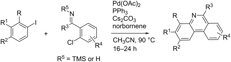
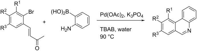
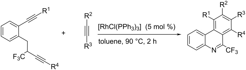
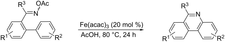
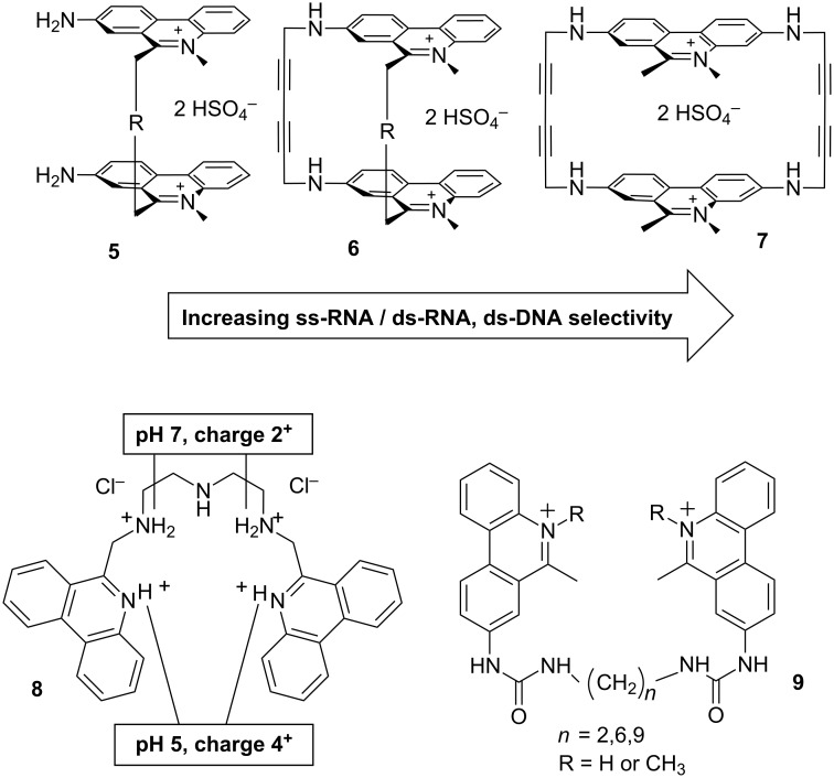
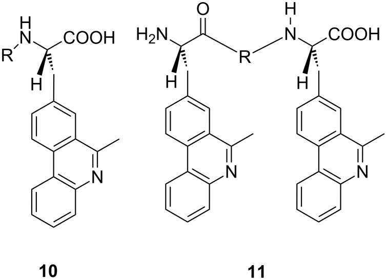
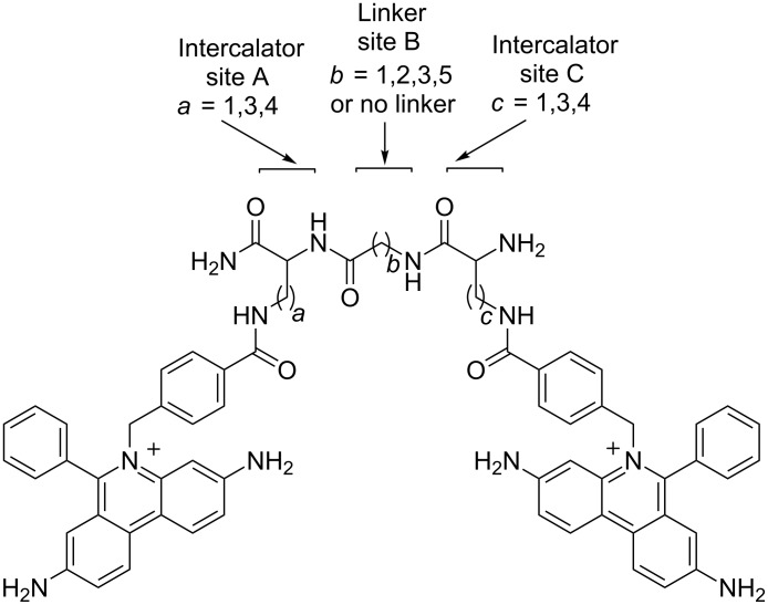
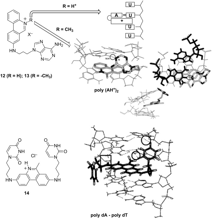
Phenanthridine derivatives are one of the most intensively studied families of biologically active compounds with efficient DNA binding capability. Attracting attention since DNA structure discovery (1960s), they were early recognized as a symbol of DNA intercalative binding, for many decades applied as gold-standard DNA- and RNA-fluorescent markers (ethidium bromide), probes for cell viability (propidium iodide), but also "ill-famed" for various toxic (genotoxic) and mutagenic effects. After two decades of low interest, the discovery of phenanthridine alkaloids and new studies of antiparasitic/antitumor properties of phenanthridine derivatives resulted in the strong increase of the scientific interest about the turn of this century. Here are summarized phenanthridine-related advances in the 21st century (2000-present period) with emphasis on the supramolecular interactions and bioorganic chemistry, as well as novel or improved synthetic approaches.

Keywords: ds-DNA and ds-RNA binding; intercalation; minor groove binding; nucleic acids; organic synthesis; phenanthridine; phenanthridinium.


Figures

Scheme 1
Scheme 1
The Grignard-based synthesis of 6-alkyl phenanthridine.
Scheme 2
Scheme 2
Radical-mediated synthesis of 6-arylphenanthridine [14].
Scheme 3
Scheme 3
A t-BuO radical-assisted homolytic aromatic substitution mechanism proposed for the conversion of diarylimine into the 6-arylphenanthridine derivatives [16].
Scheme 4
Scheme 4
Synthesis of 5,6-unsubstituted phenanthridine starting from 2-iodobenzyl chloride and aniline [17].
Scheme 5
Scheme 5
Phenanthridine synthesis initiated by UV-light irradiation photolysis of acetophenone O-ethoxycarbonyloxime derivatives at room temperature [18].
Scheme 6
Scheme 6
PhI(OAc)2-mediated oxidative cyclization of 2-isocyanobiphenyls with CF3SiMe3 [–20].
Scheme 7
Scheme 7
Targeting 6-perfluoroalkylphenanthridines [–22].
Scheme 8
Scheme 8
Easily accessible biphenyl isocyanides reacting under mild conditions (room temp., visible light irradiation, blue LED light source, N2, DMF, 10 h) with various common alkyl bromides by application the two-role catalyst [fac-Ir(ppy)3], gave phenanthridines in good yields [25].
Scheme 9
Scheme 9
Microwave irradiation of Diels–Alder adduct followed by UV irradiation of dihydrophenanthridines yielded phenanthridines [26].
Scheme 10
Scheme 10
A representative palladium catalytic cycle.
Scheme 11
Scheme 11
The common Pd-catalyst for the biphenyl conjugation results simultaneously in picolinamide-directed cyclisation; obtained N-picolinamide dihydrophenanthridine is easily converted to phenanthridine [32].
Scheme 12
Scheme 12
Pd(0)-mediated cyclisation of imidoyl-selenides forming 6-arylphenanthridine derivatives [16]. The insertion of the Pd(0) species into the carbon selenium bond followed by fast rearomatisation to phenanthridine is involved with the loss of HPdSePh.
Scheme 13
Scheme 13
Palladium-catalysed phenanthridine synthesis.
Scheme 14
Scheme 14
Aerobic domino Suzuki coupling combined with Michael addition reaction in the presence of a Pd(OAc)2/K3PO4 catalytic system in water [–35].
Scheme 15
Scheme 15
Rhodium-catalysed alkyne [2 + 2 + 2] cycloaddition reactions [36].
Scheme 16
Scheme 16
The O-acetyloximes derived from 2′-arylacetophenones underwent N–O bond cleavage and intramolecular N-arylation, followed by cross-coupling or directed C–H arylation [37].
Scheme 17
Scheme 17
C–H arylation with aryl chloride in the presence of a simple diol complex with KOt-Bu (top) [39]; for some cases it worked also in the absence of diol (bottom) [40].
Scheme 18
Scheme 18
The subsequent aza-Claisen rearrangement, ring-closing enyne metathesis and Diels–Alder reaction – a new “three-atom economic process” of phenanthridine synthesis [41].
Scheme 19
Scheme 19
Phenanthridine central-ring cyclisation with simultaneous radical-driven phosphorylation [42].
Scheme 20
Scheme 20
Three component reaction yielding the benzo[a]phenanthridine core in excellent yields [44].
Scheme 21
Scheme 21
a) Reaction of malononitrile and 1,3-indandione with BEP to form the cyclised DPP products; b) pH controlled reversible cyclisation process of DPP compounds [45].
Figure 1
Figure 1
Schematic presentation of the intercalative binding mode by the neighbour exclusion principle and important structural features of ethidium bromide: A) amino substituents responsible for fluorescence increase upon DNA intercalation; B) phenyl substituent for steric control and also impact on fluorimetric properties; C) permanent positive charge for aqueous solubility and electrostatic attraction to the DNA or RNA phosphate backbone.
Figure 2
Figure 2
Urea and guanidine derivatives of EB with modified DNA interactions [57].
Figure 3
Figure 3
Structure of mono- (3) and bis-biguanide (4) derivative. Fluorescence (y-axis normalised to starting fluorescence of free 4, c = 1.0 × 10⁻⁶ mol dm−3) was quenched by GC-DNA and increased for AT-DNA. Inset: induced (I)CD spectra λ > 280 nm of 4 (r(4)/DNA = 0.3; c(DNA) = 2 × 10⁻⁵ mol dm−3) – strong positive ICD band for AT-DNA and negative ICD band for GC-DNA. Adapted with permission from [58]. Copyright 2011 The Royal Society of Chemistry.
Scheme 22
Scheme 22
Bis-phenanthridinium derivatives (57; inert aliphatic linkers, R = –(CH2)4– or –(CH2)6–): rigidity of a “cage” – steric control of binding site. Triamine-linked bis-phenanthridine 8, note reversible doubling of positive charges at pH 5 in respect to neutral conditions (pH 7). Bis-urea phenanthridines (general structure 9): different from amino analogues (5) by fluorimetric response and DNA- and RNA-binding modes.
Figure 4
Figure 4
Series of amino acid–phenanthridine building blocks (general structure 10; R = H; Gly) and peptide-bridged bis-phenanthridine derivatives (general structure 11; R = X; Gly; Gly–Gly) [68].
Figure 5
Figure 5
General structure of 45 bis-ethidium bromide analogues. Reproduced with permission from [69]. Copyright 2012 Elsevier Ltd.
Scheme 23
Scheme 23
Top: Recognition of poly(U) by 12 and ds-polyAH+ by 13; bottom: Recognition of poly(dA)–poly(dT) by 14, intramolecular H-bonds marked by circles. Reproduced with permission from [–73], copyright 2002, 2005 The Royal Society of Chemistry and with permission from [71], copyright 2003 John Wiley & Sons, Inc.
Figure 6
Figure 6
The bis-phenanthridinium–adenine derivative 15 (LEFT) showed selectivity towards complementary UMP; structure of the 15–UMP complex (RIGHT) obtained by molecular modelling. Reproduced with permission from [75]. Copyright 2010 Elsevier Ltd.
Figure 7
Figure 7
The neomycin–methidium conjugate targeting DNA:RNA hybrid structures [80].
Figure 8
Figure 8
Two-colour RNA intercalating probe for cell imaging applications: Left: Chemical structure of EB-fluorescein conjugate (FLEth) and cartoon depicting the energy transfer process from fluorescein to the intercalated phenanthridine fluorophore. Reproduced with permission from [83]. Copyright 2008 American Chemical Society.; Right: Convenient Reporter for Small Interfering RNAs fluorophore. Reproduced with permission from [84]. Copyright 2009 American Chemical Society.
Figure 9
Figure 9
The ethidium bromide nucleosides 17 (top) and 18 (bottom). DNA duplex set 1 and 2 (E = phenanthridinium intercalation site). Reproduced with permission from [87]. Copyright 2004 American Chemical Society.
Figure 10
Figure 10
Left: various DNA duplexes; DNA1 and DNA2 used to study the impact on the adjacent basepair type on the EB fluorescence (reproduced with permission from [90], copyright 2004 John Wiley & Sons, Inc.) and DNA1,2,3,4-XY studying the EB fluorescence quenching by 7-deazaguanine (B) as a function of different position of abasic site (S). Reproduced with permission from [91] Copyright2005 Royal Society of Chemistry. Right: structure of incorporated EB (18) and of the abasic site (S).
Figure 11
Figure 11
Structure of 4,9-DAP derivative 19; Rright: MIAPaCa-2 cells stained with 10 μM 19 after 60 and 120 min incubation, respectively. Magnification 630×. Reproduced with permission from [95]. Copyright 2000 Royal Society of Chemistry.
Figure 12
Figure 12
Examples of naturally occurring phenanthridine analogues.

References

    1. Browning C H, Morgan G T, Robb J V M, Walls L P. J Pathol Bacteriol. 1938;46:203–204. doi: 10.1002/path.1700460121. - DOI
    1. Nishiwaki H, Miura M, Imai K, Ono R, Kawashima K. Cancer Res. 1974;34:2699–2703. - PubMed
    1. Parenty A D C, Smith L V, Guthrie K M, Long D-L, Plumb J, Brown R, Cronin L. J Med Chem. 2005;48:4504–4506. doi: 10.1021/jm050320z. - DOI - PubMed
    1. Kock I, Heber D, Weide M, Wolschendorf U, Clement B. J Med Chem. 2005;48:2772–2777. doi: 10.1021/jm0490888. - DOI - PubMed
    1. Krasiński A, Radić Z, Manetsch R, Raushel J, Taylor P, Sharpless K B, Kolb H C. J Am Chem Soc. 2005;127:6686–6692. doi: 10.1021/ja043031t. - DOI - PubMed