. 2014 Oct 15;7(11):7699-707.
eCollection 2014.

A key mediator, PTX3, of IKK/IκB/NF-κB exacerbates human umbilical vein endothelial cell injury and dysfunction

Affiliations

A key mediator, PTX3, of IKK/IκB/NF-κB exacerbates human umbilical vein endothelial cell injury and dysfunction

Yongbo Zhao et al. Int J Clin Exp Pathol. .

Abstract

Objective: This study was performed to investigate PTX3-mediated iNOS expression and IKK/IκB/NF-κB activation in PA-induced atherosclerotic HUVECs injury model.

Methods: The cell viability was detected by the CCK8 assay. The cell apoptosis was assessed by annexin V-PI double-labeling staining. Expression of genes and proteins were analyzed by real-time PCR and western blotting respectively. Cells were transfected with siRNAs as a gene silencing methods.

Results: PA induced cell apoptosis in human umbilical vein endothelial cells in a time and dose-dependent manner. PA also induced upregulation expression of PTX3. TPCA-1, an inhibitor of IKK-2, could suppress the expression of PTX3 and phospho-IκB-α in PA-induced endothelial dysfunction cell model. We also found that transfection of cells with PTX3 siRNA reduced the expression of iNOS and NO, and protected PA-induced cell apoptosis in HUVECs.

Conclusions: PTX3 could exacerbate endothelial dysfunction, at least partially, through IKK/IκB/NF-κB activation and overexpression of iNOS and NO, and advance the development of atherosclerosis.

Keywords: HUVECs; NF-κB; PTX3; atherosclerosis; iNOS.


Figures

Figure 1
Figure 1
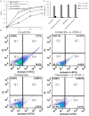
Palmitic acid-induced the apoptosis of human umbilical vein endothelial cells (HUVECs). HUVECs were incubated with various concentrations of Palmitic acid (PA) for 24 h, 48 h or 72 h, and the cell viability was examined by CCK8 assay (A). Cells were treated with vehicle, 0.2 mM PA, 0.4 mM PA or 0.8 mM PA for 48 h, the percentage of apoptotic cells was also analyzed by flow cytometric analysis of annexin V/PI double staining (B). mRNA and protein expression of pentraxin3 (PTX3) in HUVECs. Cells were treated with PA (80 mM) for 48 h. mRNA (C) and protein (C and D) expression were measured by Quantitative real-time PCR and western blotting respectively. Values are expressed as mean ± SEM, n=3 in each group. *P < 0.05, versus untreatment group.
Figure 2
Figure 2
Screening an inhibitor of I kappa B kinase-2 (IKK-2) library and validation functions of TPCA-1. HUVECs were adopted with PA (80 mM) for 48 h, made as the atherosclerotic endothelial cell injury model. Cells were treated with vehicle or inhibitor of IKK-2 (30 μM) for 48 h, followed centrifugation to obtain the supernatant. PTX-3 levels were measured by the ELISA assay (A). HUVECs were treated with untreatment, TPCA-1 only, 0.8 mM PA only and 0.8 mM PA plus TPCA-1 for 48 h, the mRNA (B) and protein (B and C) expression were measured by Quantitative real-time PCR and western blotting respectively, and the protein expression of phospho-IκB-α and IκB-α were measured by western blotting (D and E). Values are expressed as mean ± SEM, n=3 in each group. *P < 0.05, versus untreatment group.
Figure 3
Figure 3
The small interfering RNA for suppressing the function of PTX3 (si-PTX3). Three different small interfering RNA were transfected into HUVECs suppressing the mRNA expression of PTX3 (A). HUVECs were treated with untreatment, 0.8 mM PA only and 0.8 mM PA plus si-PTX3 for 48 h, the protein expression was measured by western blotting (B), and the NO concentration was detected by ELISA assay (C). Values are expressed as mean ± SEM, n=3 in each group. *P < 0.05, versus untreatment group.
Figure 4
Figure 4
PTX3-induced the apoptosis of human umbilical vein endothelial cells (HUVECs). HUVECs were treated with untreatment, si-PTX3 only, 0.8 mM PA only and 0.8 mM PA plus si-PTX3 for 48 h, and the cell viability was examined by CCK8 assay (A). The apoptotic cells were detected by flow cytometric analysis of annexin V/PI double staining (B). (C) The percntage of apoptotic cells was also analyzed by annexin V/PI double staining (n=3).

References

    1. Wahyudi S, Sargowo D. Green tea polyphenols inhibit oxidized LDL-induced NF-KB activation in human umbilical vein endothelial cells. Acta Med Indones. 2007;39:66–70. - PubMed
    1. Shi J, Sun X, Lin Y, Zou X, Li Z, Liao Y, Du M, Zhang H. Endothelial cell injury and dysfunction induced by silver nanoparticles through oxidative stress via IKK/NF-kappaB pathways. Biomaterials. 2014;35:6657–6666. - PubMed
    1. Lee CH, Lee SD, Ou HC, Lai SC, Cheng YJ. Eicosapentaenoic acid protects against palmitic acid-induced endothelial dysfunction via activation of the AMPK/eNOS pathway. Int J Mol Sci. 2014;15:10334–10349. - PMC - PubMed
    1. Khan MJ, Rizwan Alam M, Waldeck-Weiermair M, Karsten F, Groschner L, Riederer M, Hallstrom S, Rockenfeller P, Konya V, Heinemann A, Madeo F, Graier WF, Malli R. Inhibition of autophagy rescues palmitic acid-induced necroptosis of endothelial cells. J Biol Chem. 2012;287:21110–21120. - PMC - PubMed
    1. Staiger H, Staiger K, Stefan N, Wahl HG, Machicao F, Kellerer M, Haring HU. Palmitate-induced interleukin-6 expression in human coronary artery endothelial cells. Diabetes. 2004;53:3209–3216. - PubMed

MeSH terms

LinkOut - more resources