Accuracy of 3D white light scanning of abutment teeth impressions: evaluation of trueness and precision
- PMID: 25551007
- PMCID: PMC4279045
- DOI: 10.4047/jap.2014.6.6.468
Accuracy of 3D white light scanning of abutment teeth impressions: evaluation of trueness and precision
Abstract
Purpose: This study aimed to evaluate the accuracy of digitizing dental impressions of abutment teeth using a white light scanner and to compare the findings among teeth types.
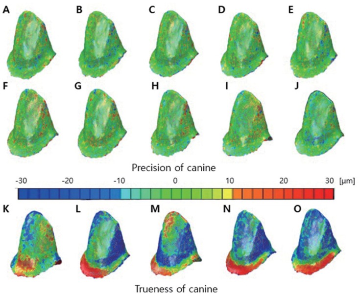
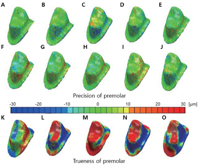
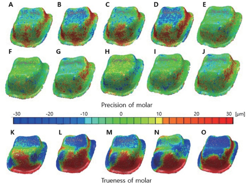
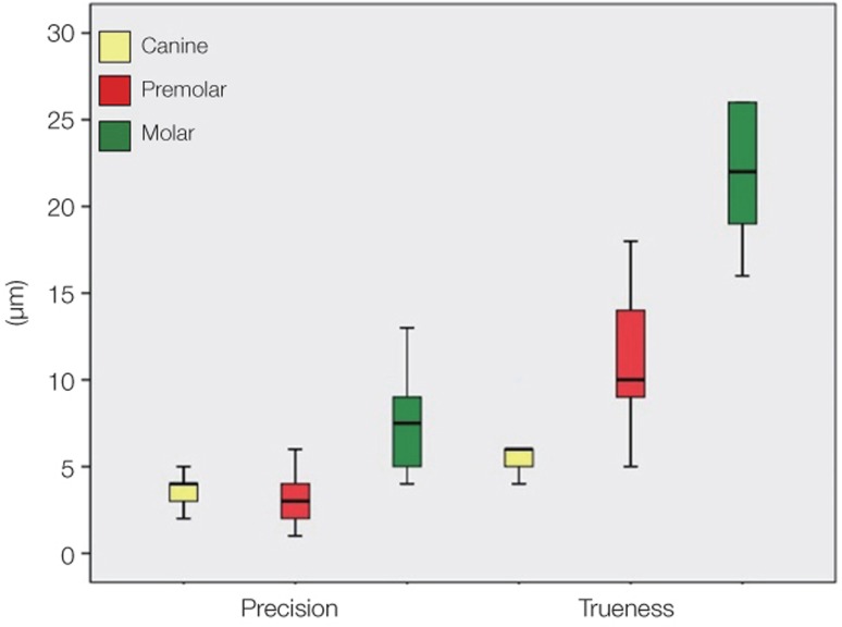
Materials and methods: To assess precision, impressions of the canine, premolar, and molar prepared to receive all-ceramic crowns were repeatedly scanned to obtain five sets of 3-D data (STL files). Point clouds were compared and error sizes were measured (n=10 per type). Next, to evaluate trueness, impressions of teeth were rotated by 10°-20° and scanned. The obtained data were compared with the first set of data for precision assessment, and the error sizes were measured (n=5 per type). The Kruskal-Wallis test was performed to evaluate precision and trueness among three teeth types, and post-hoc comparisons were performed using the Mann-Whitney U test with Bonferroni correction (α=.05).
Results: Precision discrepancies for the canine, premolar, and molar were 3.7 µm, 3.2 µm, and 7.3 µm, respectively, indicating the poorest precision for the molar (P<.001). Trueness discrepancies for teeth types were 6.2 µm, 11.2 µm, and 21.8 µm, respectively, indicating the poorest trueness for the molar (P=.007).
Conclusion: In respect to accuracy the molar showed the largest discrepancies compared with the canine and premolar. Digitizing of dental impressions of abutment teeth using a white light scanner was assessed to be a highly accurate method and provided discrepancy values in a clinically acceptable range. Further study is needed to improve digitizing performance of white light scanning in axial wall.
Keywords: 3D shape data; Accuracy; Impression scanning; Point cloud; Precision and trueness; White light scanner.
Figures
References
-
- Naidu D, Freer TJ. Validity, reliability, and reproducibility of the iOC intraoral scanner: a comparison of tooth widths and Bolton ratios. Am J Orthod Dentofacial Orthop. 2013;144:304–310. - PubMed
-
- Flügge TV, Schlager S, Nelson K, Nahles S, Metzger MC. Precision of intraoral digital dental impressions with iTero and extraoral digitization with the iTero and a model scanner. Am J Orthod Dentofacial Orthop. 2013;144:471–478. - PubMed
-
- Persson AS, Odén A, Andersson M, Sandborgh-Englund G. Digitization of simulated clinical dental impressions: virtual three-dimensional analysis of exactness. Dent Mater. 2009;25:929–936. - PubMed
-
- Quaas S, Rudolph H, Luthardt RG. Direct mechanical data acquisition of dental impressions for the manufacturing of CAD/CAM restorations. J Dent. 2007;35:903–908. - PubMed
-
- Persson A, Andersson M, Oden A, Sandborgh-Englund G. A three-dimensional evaluation of a laser scanner and a touchprobe scanner. J Prosthet Dent. 2006;95:194–200. - PubMed
LinkOut - more resources
Full Text Sources
Other Literature Sources