. 2014 Dec 2:5:2308-15.
doi: 10.3762/bjnano.5.239. eCollection 2014.

Coating with luminal gut-constituents alters adherence of nanoparticles to intestinal epithelial cells

Affiliations

Coating with luminal gut-constituents alters adherence of nanoparticles to intestinal epithelial cells

Heike Sinnecker et al. Beilstein J Nanotechnol. .

Abstract

Background: Anthropogenic nanoparticles (NPs) have found their way into many goods of everyday life. Inhalation, ingestion and skin contact are potential routes for NPs to enter the body. In particular the digestive tract with its huge absorptive surface area provides a prime gateway for NP uptake. Considering that NPs are covered by luminal gut-constituents en route through the gastrointestinal tract, we wanted to know if such modifications have an influence on the interaction between NPs and enterocytes.

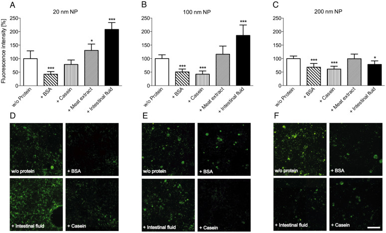
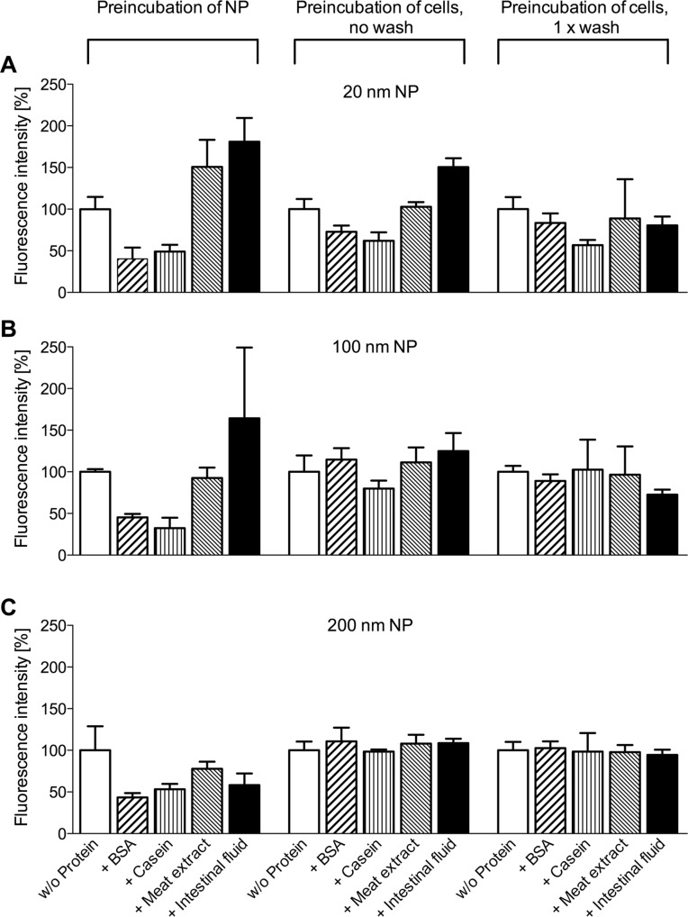
Results: We investigated the consequences of a treatment with various luminal gut-constituents on the adherence of nanoparticles to intestinal epithelial cells. Carboxylated polystyrene particles 20, 100 and 200 nm in size represented our anthropogenic NPs, and differentiated Caco-2 cells served as model for mature enterocytes of the small intestine. Pretreatment with the proteins BSA and casein consistently reduced the adherence of all NPs to the cultured enterocytes, while incubation of NPs with meat extract had no obvious effect on particle adherence. In contrast, contact with intestinal fluid appeared to increase the particle-cell interaction of 20 and 100 nm NPs.

Conclusion: Luminal gut-constituents may both attenuate and augment the adherence of NPs to cell surfaces. These effects appear to be dependent on the particle size as well as on the type of interacting protein. While some proteins will rather passivate particles towards cell attachment, possibly by increasing colloid stability or camouflaging attachment sites, certain components of intestinal fluid are capable to modify particle surfaces in such a way that interactions with cellular surface structures result in an increased binding.

Keywords: adherence; agglomeration; intestinal epithelial cells; nanoparticles (NPs); protein.


Figures

Figure 1
Figure 1
Differentiation of Caco-2 cells over 21 days post-confluence. a,b: With reaching confluence, cells have built a monolayer and rudimentary microvilli are visible; c,d: 7 days after formation of a confluent monolayer microvilli are pronounced; e,f: 14 days post-confluence cells are grown to a columnar epithelium, the microvilli have increased, tight junctions are formed and the glycocalyx between and on the top of the microvilli is visible; g,h: 21 days post-confluence all structural attributes of intestinal epithelial cells are formed. Upper panel (a,c,e,g): Scale bar, 5 µm, lower panel (b,d,f,h): Scale bar, 1 µm.
Figure 2
Figure 2
Interaction of fluorescent NPs with Caco-2 cells in the presence of proteinaceous compounds. Differently sized NPs, without or with pretreatment with different protein mixtures, were incubated with Caco-2 cells. After extensive washing, particle adherence to and/or uptake by the cells was determined by fluorescence microscopy. Values were normalized to intensities obtained with untreated particles (A, B, C; mean + SD of 6–8 individual experiments; p < 0.05 (*) and p < 0.001 (***), ANOVA with Dunnett’s post-hoc test). En-face fluorescence microscope images (D, E, F) show variations in NP adherence, depending on the pretreatment with proteins. Scale bar, 100 µm.
Figure 3
Figure 3
Adherence of 100 nm NPs to Caco-2 cells. The glycocalyx was counter-stained with a lectin (red) to visualize the cell surface of the monolayer. Stacks of 2-dimensional microscope images were processed with the software Imaris x64 to give a 3D rendition. A: In the absence of BSA, NPs are attached to the cell surface mainly in form of particle agglomerates (green spots in variable size). B: After preincubation of NPs with BSA only few and small particle spots are visible. Scale bar: 15 µm.
Figure 4
Figure 4
Interaction of fluorescent NPs with cells after preincubation with proteinaceous compounds. Either differently sized NPs or Caco-2 cells were preincubated with buffer or with different proteins before interacting with each other. Particle adherence to the cells was determined by fluorescence microscopy. Values were normalized to intensities obtained with untreated cells and particles (mean + SD of three experiments).
Figure 5
Figure 5
Schematic model of possible interaction mechanisms between differently sized NPs and endothelial cell surface. A: Non-specific binding of “naked” small NPs to outer surface structures and membrane components; B: Decreased non-specific binding of small NPs after “passivation” with proteins; C: Increased binding of small NPs pretreated with luminal gut-constituents to specific attachment sites within the glycocalyx; D: Non-specific binding of “naked” large NPs to outer surface structures of the cells; E: Abolished binding of large NPs pretreated with luminal gut-constituents since particles are too large to penetrate the glycocalyx and reach specific attachment sites.

References

    1. Chaudhry Q, Scotter M, Blackburn J, Ross B, Boxall A, Castle L, Aitken R, Watkins R. Food Addit Contam, Part A. 2008;25:241–258. doi: 10.1080/02652030701744538. - DOI - PubMed
    1. Thomas T, Thomas K, Sadrieh N, Savage N, Adair P, Bronaugh R. Toxicol Sci. 2006;91:14–19. doi: 10.1093/toxsci/kfj129. - DOI - PubMed
    1. Antonini J M, Lewis A B, Roberts J R, Whaley D A. Am J Ind Med. 2003;43:350–360. doi: 10.1002/ajim.10194. - DOI - PubMed
    1. Dennekamp M, Mehenni O H, Cherrie J W, Seaton A. Ann Occup Hyg. 2002;46(Suppl 1):412–414. doi: 10.1093/annhyg/mef702. - DOI
    1. Churg A, Wright J L. Chest. 2002;122:306S–309S. doi: 10.1378/chest.122.6_suppl.306S. - DOI - PubMed

LinkOut - more resources