A swelling of the maxilla: a case report and differential diagnosis
- PMID: 25551097
- PMCID: PMC4279968
- DOI: 10.5125/jkaoms.2014.40.6.308
A swelling of the maxilla: a case report and differential diagnosis
Abstract
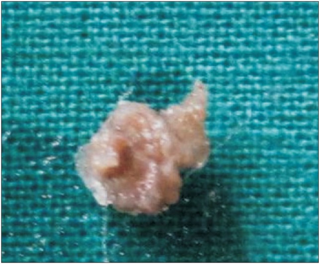
Ossifying fibromas are benign fibro-osseous tumors of mesenchymal origin. Although ossifying fibromas have principally been found in the jaw, they have also been reported in the frontal, ethmoid, sphenoid, and temporal bones, as well as the orbit and anterior cranial fossa. Ossifying fibromas affecting the jaw exhibit variable behaviors ranging from slow growth to occasionally aggressive local destruction. In the present article, we discuss a differential diagnosis considered for maxillary swellings and report a rare case of ossifying fibroma occurring in the maxilla.
Keywords: Bone neoplasms; Maxilla; Ossifying fibroma; Tomography.
Conflict of interest statement
No potential conflict of interest relevant to this article was reported.
Figures


Similar articles
-
Aggressive psammomatoid ossifying fibromas of the sinonasal region: a clinicopathologic study of a distinct group of fibro-osseous lesions.Cancer. 1995 Oct 1;76(7):1155-65. doi: 10.1002/1097-0142(19951001)76:7<1155::aid-cncr2820760710>3.0.co;2-p. Cancer. 1995. PMID: 8630892
-
Juvenile Ossifying Fibroma of Maxilla.Kathmandu Univ Med J (KUMJ). 2018 Jul-Sept.;16(63):263-265. Kathmandu Univ Med J (KUMJ). 2018. PMID: 31719318
-
An Uncommon Presentation of Ossifying Fibroma in the Maxilla.Cureus. 2022 Mar 30;14(3):e23638. doi: 10.7759/cureus.23638. eCollection 2022 Mar. Cureus. 2022. PMID: 35510009 Free PMC article.
-
Juvenile psammomatoid ossifying fibroma of maxillary sinus: case report with review of literature.J Maxillofac Oral Surg. 2014 Jun;13(2):109-14. doi: 10.1007/s12663-013-0479-6. Epub 2013 Feb 7. J Maxillofac Oral Surg. 2014. PMID: 24822000 Free PMC article. Review.
-
Central ossifying fibroma of the anterior maxilla: report of case.J Am Dent Assoc. 1988 Apr;116(4):507-10. doi: 10.14219/jada.archive.1988.0304. J Am Dent Assoc. 1988. PMID: 3288667 Review.
Cited by
-
Multidisciplinary approach for management of a patient with fibrous dysplasia of maxilla.BMJ Case Rep. 2015 Aug 5;2015:bcr2015210330. doi: 10.1136/bcr-2015-210330. BMJ Case Rep. 2015. PMID: 26245286 Free PMC article.
-
Craniomaxillofacial Fibrous Dysplasia Improved Cosmetic and Occlusal Problem by Comprehensive Treatment: A Case Report and Review of Current Treatments.Diagnostics (Basel). 2022 Sep 3;12(9):2146. doi: 10.3390/diagnostics12092146. Diagnostics (Basel). 2022. PMID: 36140547 Free PMC article.
References
-
- Gondivkar SM, Gadbail AR, Chole R, Parikh RV, Balsaraf S. Ossifying fibroma of the jaws: report of two cases and literature review. Oral Oncol. 2011;47:804–809. - PubMed
-
- Waldron CA. Fibro-osseous lesions of the jaws. J Oral Maxillofac Surg. 1993;51:828–835. - PubMed
-
- Wood NK, Goaz PW. Differential diagnosis of oral and maxillofacial lesions. 5th ed. St. Loius: Mosby; 1998.
-
- White SC, Pharoah MJ. Diseases of bone manifested in the jaws. In: White SC, Pharoah MJ, editors. Oral radiology: principles and interpretation. 5th ed. St. Loius: Mosby; 2004. p. 412.
-
- Orpe EC, Lee L, Pharoah MJ. A radiological analysis of chronic sclerosing osteomyelitis of the mandible. Dentomaxillofac Radiol. 1996;25:125–129. - PubMed
Publication types
LinkOut - more resources
Full Text Sources
Other Literature Sources