Removal of well-fixed components in femoral revision arthroplasty with controlled segmentation of the proximal femur
- PMID: 25551372
- PMCID: PMC4299768
- DOI: 10.1186/s13018-014-0137-9
Removal of well-fixed components in femoral revision arthroplasty with controlled segmentation of the proximal femur
Abstract
Background: The transfemoral and the extended trochanteric osteotomies are the most common osteotomies used in femoral revision, both when proximal or diaphyseal fixation of the new component has been decided. We present an alternative approach to the trochanteric osteotomies, most frequently used with distally fixated stems, to overcome their shortcomings of osteotomy migration and nonunion, but, most of all, the uncontrollable fragmentation of the femur.
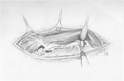
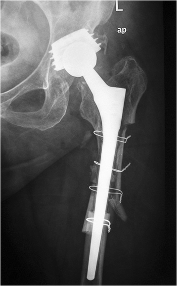
Methods: The procedure includes a complete circular femoral osteotomy just below the stem tip to prevent distal fracture propagation and a subsequent preplanned segmentation of the proximal femur for better exposure and fast removal of the old prosthesis. The bone fragments are reattached with cerclage wires to the revision prosthesis, which is safely anchored distally. A modified posterolateral approach is used, as the preservation of the continuity of the abductors, the greater trochanter, and the vastus lateralis is a prerequisite.
Results: Between 2006 and 2012, 47 stems (33 women, 14 men, mean age 68 years, range 39-88 years) were revised using this technique. They were 12 (26%) stable and 35 (74%) loose prostheses and were all revised to tapered, fluted, grit-blasted stems. No fracture of the trochanters or the distal femur occurred intraoperatively. Mean follow-up was 28 months (range 6-70 months). No case of trochanteric migration or nonunion of the osteotomies was recorded. Restoration of the preexisting bone defects occurred in 83% of the patients. Three patients required repeat revision due to dislocation and one due to a postoperative periprosthetic fracture. None of the failures was attributed to the procedure itself.
Conclusions: This new osteotomy technique may seem aggressive at first, but, at least in our hands, has effectively increased the speed of the femoral revision, particularly for the most difficult well-fixed components, but not at the expense of safety.
Figures


Similar articles
-
Extended Trochanteric Osteotomy in Revision Total Hip Arthroplasty.JBJS Essent Surg Tech. 2023 Jul 21;13(3):e21.00003. doi: 10.2106/JBJS.ST.21.00003. eCollection 2023 Jul-Sep. JBJS Essent Surg Tech. 2023. PMID: 38282724 Free PMC article.
-
Modified transfemoral approach to revision arthroplasty with uncemented modular revision stems.Oper Orthop Traumatol. 2007 Mar;19(1):32-55. doi: 10.1007/s00064-007-1194-6. Oper Orthop Traumatol. 2007. PMID: 17345026 Clinical Trial.
-
Extended trochanteric osteotomy via the direct lateral approach in revision hip arthroplasty.Clin Orthop Relat Res. 2003 Dec;(417):210-6. doi: 10.1097/01.blo.0000096818.67494.7b. Clin Orthop Relat Res. 2003. PMID: 14646719
-
Trochanteric osteotomies in revision total hip arthroplasty: contemporary techniques and results.Instr Course Lect. 2005;54:143-55. Instr Course Lect. 2005. PMID: 15948441 Review.
-
Extended trochanteric osteotomy: planning, surgical technique, and pitfalls.Instr Course Lect. 2004;53:119-30. Instr Course Lect. 2004. PMID: 15116606 Review.
Cited by
-
Development of the Revision Hip Complexity Classification using a modified Delphi technique.Bone Jt Open. 2022 May;3(5):423-431. doi: 10.1302/2633-1462.35.BJO-2022-0022.R1. Bone Jt Open. 2022. PMID: 35549448 Free PMC article.
-
Mid-term results of revision total hip arthroplasty with an uncemented modular femoral component.Hip Int. 2018 Jan;28(1):84-89. doi: 10.5301/hipint.5000522. Hip Int. 2018. PMID: 29027190 Free PMC article.
-
Trochanteric osteotomy in revision total hip arthroplasty.EFORT Open Rev. 2020 Sep 10;5(8):477-485. doi: 10.1302/2058-5241.5.190063. eCollection 2020 Aug. EFORT Open Rev. 2020. PMID: 32953133 Free PMC article. Review.
-
Treatment of Prosthetic Joint Infection due to Listeria Monocytogenes. A Comprehensive Literature Review and a Case of Total Hip Arthroplasty Infection.Arthroplast Today. 2021 Dec 13;13:48-54. doi: 10.1016/j.artd.2021.10.016. eCollection 2022 Feb. Arthroplast Today. 2021. PMID: 34977306 Free PMC article.
-
Eradication of infection, survival, and radiological results of uncemented revision stems in infected total hip arthroplasties.Acta Orthop. 2016 Dec;87(6):637-643. doi: 10.1080/17453674.2016.1237423. Epub 2016 Sep 23. Acta Orthop. 2016. PMID: 27658856 Free PMC article.
References
-
- Wagner H. Revision prosthesis for the hip joint in severe bone loss. Orthopade. 1987;16:295–300. - PubMed
-
- Böhm P, Bischel O. Femoral revision with the Wagner SL revision stem: evaluation of one hundred and twenty-nine revisions followed for a mean of 4.8 years. J Bone Joint Surg Am. 2001;83-A(7):1023–1031. - PubMed
MeSH terms
LinkOut - more resources
Full Text Sources
Other Literature Sources
Medical
Miscellaneous

