

Impairment of myocardial mitochondria in viral myocardial disease and its reflective window in peripheral cells
- PMID: 25551390
- PMCID: PMC4281208
- DOI: 10.1371/journal.pone.0116239
Impairment of myocardial mitochondria in viral myocardial disease and its reflective window in peripheral cells
Abstract
Background: Viral myocardial disease (VMD) is a common disease inducing heart failure. It has not been clear the roles of mitochondrial damage in the pathological changes of cardiomyocytes in VMD.
Methods: Myocardial tissues and lymphocytes were collected from 83 VMD patients. Control groups included 12 cases of healthy accidental death with myocardial autopsy and 23 healthy blood donors. The mouse model of viral myocarditis (VMC) was established by Coxsackie virus B3 infection and myocardial tissues and skeletal muscle were collected. Mitochondrial DNA (mtDNA) deletion rate was quantitatively determined using polymerase chain reaction.
Results: There was significantly difference of myocardial mitochondrial DNA deletion rate between VMD or VMC group and control group (P<0.05). Moreover, the loss of mitochondrial membrane phospholipids was significantly different between VMD or VMC group and control group. In VMC mice, there were negative correlations between myocardial mtDNA3867 deletion rate and left ventricular peak systolic pressure (LVPSP) (r = -0.66, P<0.05), and between myocardial mtDNA3867 deletion rate and +dp/dtmax (r = -0.79, P<0.05), while there was positive correlation between myocardial mtDNA3867 deletion rate and -dp/dtmax (r = 0.80, P<0.05).
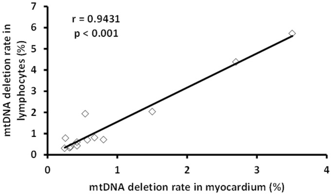
Conclusion: Mitochondrial damage is an important pathophysiological mechanism leading to myocardial injury and cardiac dysfunction. The mitochondrial damage in the skeletal muscle and lymphocytes reflect a "window" of myocardial mitochondrial damage.
Conflict of interest statement
Figures


Similar articles
-
Impairment of myocardial and skeletal mitochondria in mice with viral myocarditis and their correlation.J Huazhong Univ Sci Technolog Med Sci. 2007 Jun;27(3):237-40. doi: 10.1007/s11596-007-0305-9. J Huazhong Univ Sci Technolog Med Sci. 2007. PMID: 17641831
-
Myocardial Mitochondrial DNA Drives Macrophage Inflammatory Response through STING Signaling in Coxsackievirus B3-Induced Viral Myocarditis.Cells. 2023 Oct 31;12(21):2555. doi: 10.3390/cells12212555. Cells. 2023. PMID: 37947632 Free PMC article.
-
Inhibition of Drp1 attenuates mitochondrial damage and myocardial injury in Coxsackievirus B3 induced myocarditis.Biochem Biophys Res Commun. 2017 Mar 11;484(3):550-556. doi: 10.1016/j.bbrc.2017.01.116. Epub 2017 Jan 25. Biochem Biophys Res Commun. 2017. PMID: 28131843
-
Electron transport chain defects in heart failure.Heart Fail Rev. 2002 Apr;7(2):131-9. doi: 10.1023/a:1015372407647. Heart Fail Rev. 2002. PMID: 11988637 Review.
-
Pathogenesis of myocardial injury in myocarditis and cardiomyopathy.Jpn Circ J. 1991 Nov;55(11):1132-7. doi: 10.1253/jcj.55.1132. Jpn Circ J. 1991. PMID: 1660942 Review.
Cited by
-
Mitochondria Dysfunction at the Heart of Viral Myocarditis: Mechanistic Insights and Therapeutic Implications.Viruses. 2023 Jan 26;15(2):351. doi: 10.3390/v15020351. Viruses. 2023. PMID: 36851568 Free PMC article. Review.
-
Ultrastructural Changes in Mitochondria in Patients with Dilated Cardiomyopathy and Parvovirus B19 Detected in Heart Tissue without Myocarditis.J Pers Med. 2022 Jan 28;12(2):177. doi: 10.3390/jpm12020177. J Pers Med. 2022. PMID: 35207664 Free PMC article.
-
Calumenin relieves cardiac injury by inhibiting ERS-initiated apoptosis during viral myocarditis.Int J Clin Exp Pathol. 2017 Jul 1;10(7):7277-7284. eCollection 2017. Int J Clin Exp Pathol. 2017. PMID: 31966567 Free PMC article. Review.
-
SARS-CoV-2: characteristics and current advances in research.Virol J. 2020 Jul 29;17(1):117. doi: 10.1186/s12985-020-01369-z. Virol J. 2020. PMID: 32727485 Free PMC article. Review.
-
Molecular Mechanisms behind Persistent Presence of Parvovirus B19 in Human Dilated Myocardium.Adv Exp Med Biol. 2022;1376:181-202. doi: 10.1007/5584_2021_702. Adv Exp Med Biol. 2022. PMID: 35025080
References
-
- Lv S, Rong J, Ren S, Wu M, Li M, et al. (2013) Epidemiology and diagnosis of viral myocarditis. Hellenic J Cardiol 54:382–391. - PubMed
-
- Maron BJ, Towbin JA, Thiene G, Antzelevitch C, Corrado D, et al. (2006) Contemporary definitions and classification of the cardiomyopathies: an American Heart Association Scientific Statement from the Council on Clinical Cardiology, Heart Failure and Transplantation Committee; Quality of Care and Outcomes Research and Functional Genomics and Translational Biology Interdisciplinary Working Groups; and Council on Epidemiology and Prevention. Circulation 113:1807–1816. - PubMed
-
- Caforio AL, Baboonian C, McKenna WJ (1997) Postviral autoimmune heart disease-fact or fiction. Eur Heart J 18:1051–1055. - PubMed
Publication types
MeSH terms
Substances
LinkOut - more resources
Full Text Sources
Other Literature Sources
Medical
