. 2014;10(12):2251-68.
doi: 10.4161/15548627.2014.981913.

Multiple functions of the SNARE protein Snap29 in autophagy, endocytic, and exocytic trafficking during epithelial formation in Drosophila

Affiliations

Multiple functions of the SNARE protein Snap29 in autophagy, endocytic, and exocytic trafficking during epithelial formation in Drosophila

Elena Morelli et al. Autophagy. 2014.

Abstract

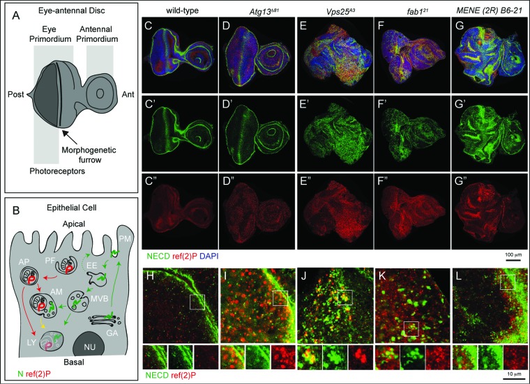
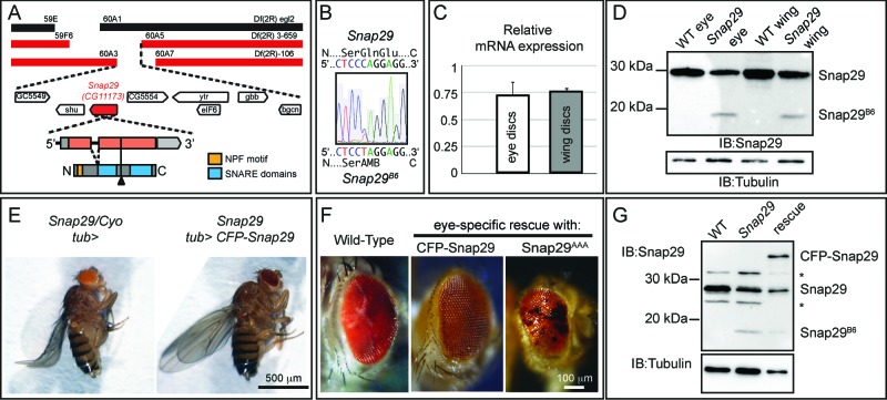
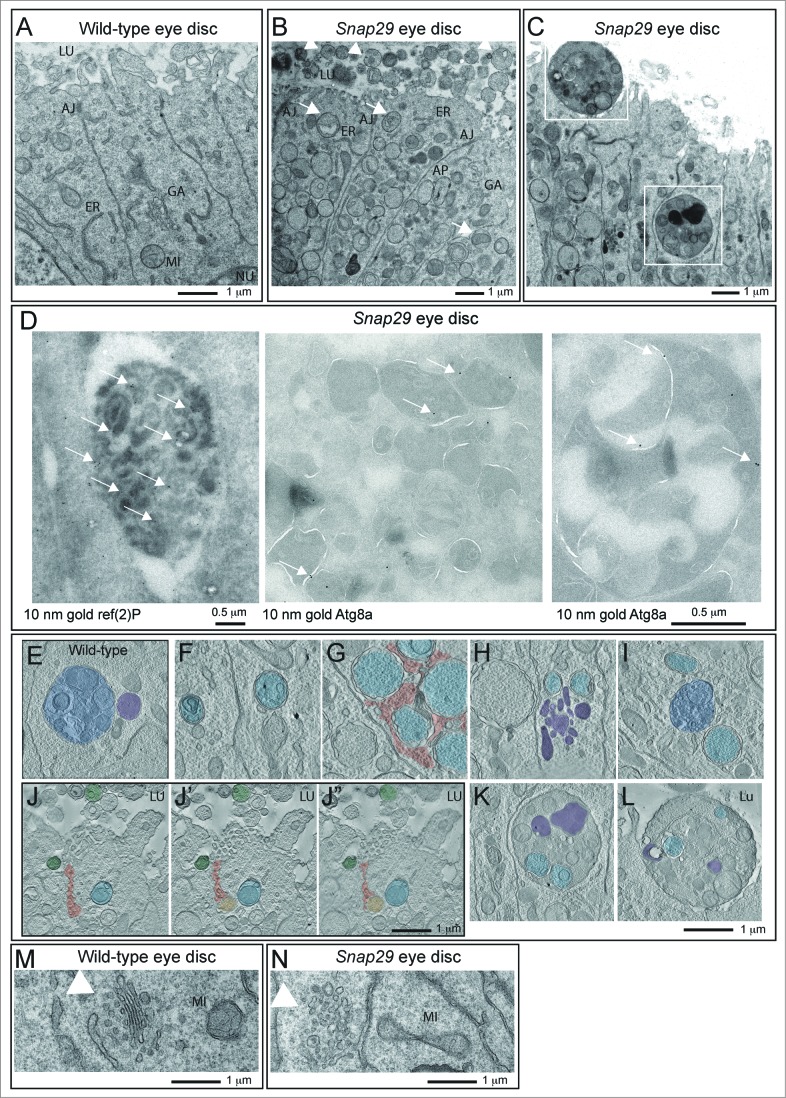
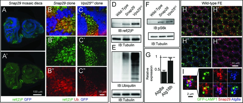
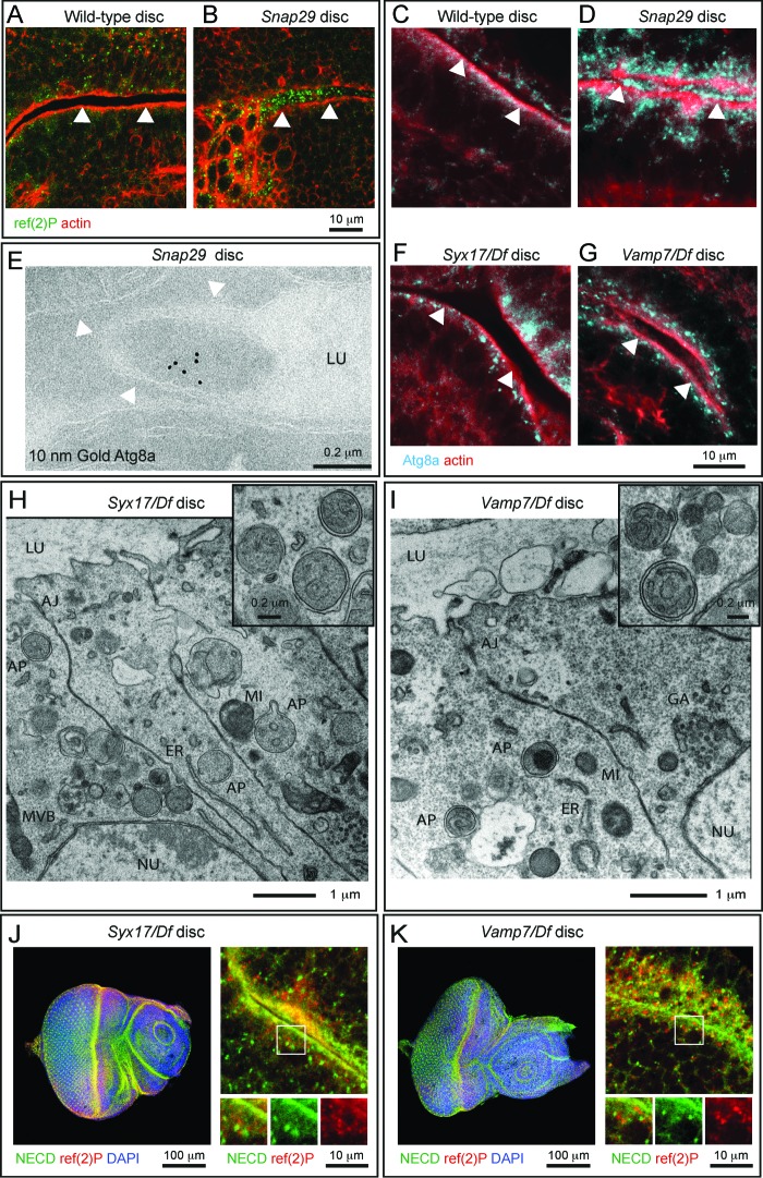
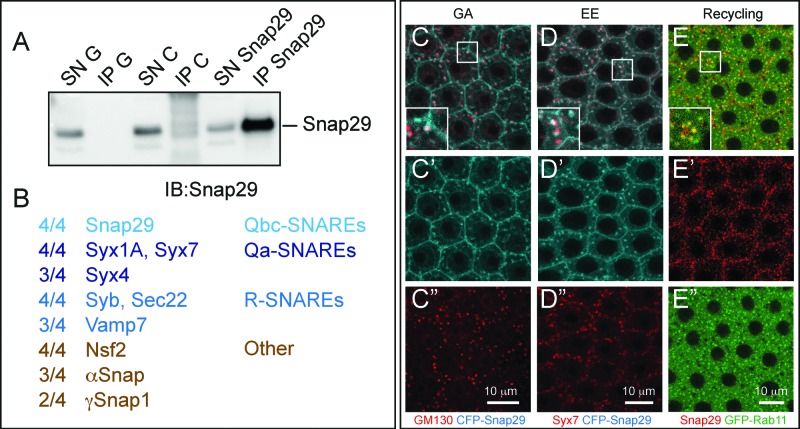
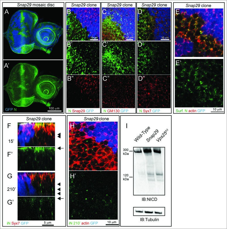
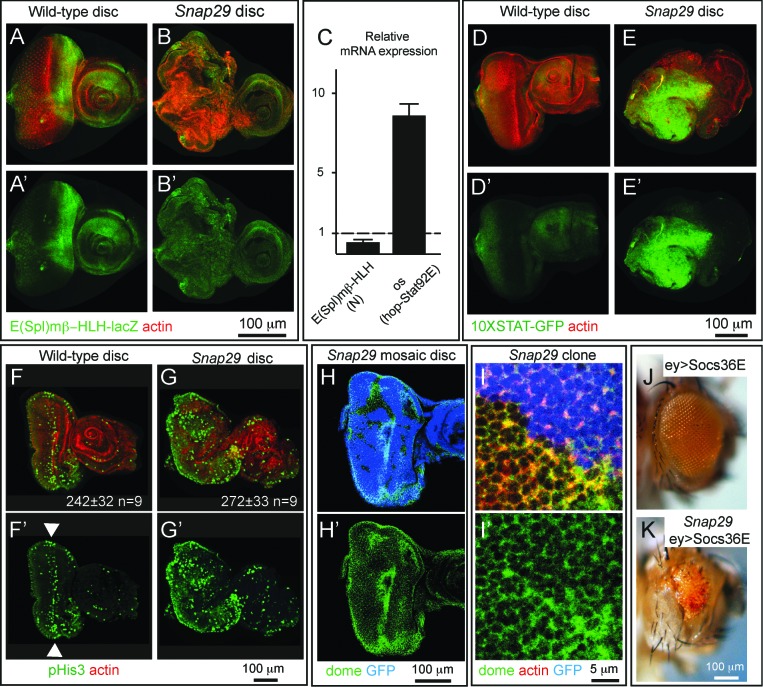
How autophagic degradation is linked to endosomal trafficking routes is little known. Here we screened a collection of uncharacterized Drosophila mutants affecting membrane transport to identify new genes that also have a role in autophagy. We isolated a loss of function mutant in Snap29 (Synaptosomal-associated protein 29 kDa), the gene encoding the Drosophila homolog of the human protein SNAP29 and have characterized its function in vivo. Snap29 contains 2 soluble NSF attachment protein receptor (SNARE) domains and a asparagine-proline-phenylalanine (NPF motif) at its N terminus and rescue experiments indicate that both SNARE domains are required for function, whereas the NPF motif is in part dispensable. We find that Snap29 interacts with SNARE proteins, localizes to multiple trafficking organelles, and is required for protein trafficking and for proper Golgi apparatus morphology. Developing tissue lacking Snap29 displays distinctive epithelial architecture defects and accumulates large amounts of autophagosomes, highlighting a major role of Snap29 in autophagy and secretion. Mutants for autophagy genes do not display epithelial architecture or secretion defects, suggesting that the these alterations of the Snap29 mutant are unlikely to be caused by the impairment of autophagy. In contrast, we find evidence of elevated levels of hop-Stat92E (hopscotch-signal transducer and activator of transcription protein at 92E) ligand, receptor, and associated signaling, which might underlie the epithelial defects. In summary, our findings support a role of Snap29 at key steps of membrane trafficking, and predict that signaling defects may contribute to the pathogenesis of cerebral dysgenesis, neuropathy, ichthyosis, and palmoplantar keratoderma (CEDNIK), a human congenital syndrome due to loss of Snap29.

Keywords: Atg, autophagy-related; CEDNIK, cerebral dysgenesis, neuropathy, ichthyosis, and palmoplantar keratoderma; CFP, cyan fluorescent protein; E(spl)mβ-HLH, enhancer of split mβ, helix-loop-helix; EM, electron microscopy; ESCRT, endosomal sorting complex required for transport; FE, follicular epithelium; GFP, green fluorescent protein; MENE, mutant eye no eclosion; MVB, multivesicular body; N, Notch; NECD, N extracellular domain; NPF, asparagine-proline-phenylalanine; Notch; SNARE; SNARE, soluble NSF attachment protein receptor; Snap29; Snap29, synaptosomal-associated protein 29 kDa; Socs36E, suppressor of cytokine signaling at 36E; Syb, Synaptobrevin; Syx, syntaxin; V-ATPase, vacuolar H+-ATPase; Vamp, vesicle-associated membrane protein; Vps25, vacuolar protein sorting 25; WT, wild type; autophagy; dome; dome, domeless; histone H3, His3; hop-Stat92E, hopscotch-signal transducer and activator of transcription protein at 92E; os, outstretched; ref(2)P, refractory to sigma P; trafficking; usnp.


Figures

Figure 1.
Figure 1.
An assay to identify novel trafficking genes involved in autophagy. (A and B) Schematic view of subapical cross-section through a third-instar larval eye-antennal imaginal discs (A) and of an epithelial cell contained in it (B). B depicts the secretion and endocytic degradation routes followed by N, and the autophagic degradation routes highlighted by ref(2)P. AM, amphisome; Ant, anterior; AP, autophagosome; EE, early endosome; GA, Golgi apparatus; LY, lysosome; NU, Nucleus; PG, phagophore; PM, plasma membrane; Post, posterior. (C to G) WT and mutant discs of the indicated genotype immunostained to detect N, ref(2)p and nuclei. Ato E″ show the N and ref(2)P channels, respectively. Compared to WT, discs mutant for the Atg13 display strong accumulation of ref(2)P, no N accumulation and normal organ morphology. In Vps25 and fab1 mutant discs N and ref(2)P accumulate, however only in Vps25 mutant discs disc morphology is aberrant. MENE (2R)-E B6 mutant discs display epithelial architecture defects, and strong ref(2)P and N accumulation. (H to L) High magnification of a cross-section of the anterior portion of mutant discs, as in C to G. Enlargements of the boxed area and its single channels are shown below each panel. Note the distinct patterns of accumulation of N, or ref(2)P, in the different mutants.
Figure 2.
Figure 2.
MENE(2R)-E B6 is a null mutant of Drosophila Snap29. (A) Schematic view of the Snap29 locus. Df(2R)egl2 (black) complements the B6-21 mutation, while Df(2R)3-659 and Df(2R)106 (red) fail to complement it, indicating that B6-21 maps to the genetic interval 60A3-A5 on the right arm of the Drosophila chromosome 2. The coding sequence of Snap29 is shown in orange, while the domains of Snap29 are indicated in yellow and blue. A black triangle marks the approximate position of the B6 mutation. (B) Sequencing of the B6 allele in heterozygosity with the parental chromosome on which the mutation was induced. A C-to-T change creates a premature stop codon that truncates the protein right after the first SNARE domain. (C) Expression of Snap29 mRNA is only 25% reduced in mutant eye-antennal and wing discs, relative to WT. (D) Analysis of Snap29 expression by protein gel blot in WT disc extracts and in extracts of discs containing Snap29B6 mutant cells indicates that Snap29B6, a truncated form of Snap29, is present in mutant cells. (E) Ubiquitous expression of CFP-Snap29 under tubulin-Gal4 (tub>) rescues lethality of homozygous Snap29 flies. Rescued flies (right) are indistinguishable from heterozygous animals (left). (F) Adult eyes of flies with the indicated genetic background. Eye-specific ectopic expression of CFP-Snap29, or of a Snap29 form with a mutated NPF motif (Snap29AAA) rescue defects of Snap29B6 mutant eye discs and yield adults with normal (CFP-Snap29), or reduced eyes (Snap29AAA). Mutant cells expressing the rescue construct give rise to orange photoreceptors, while the WT cells give rise to dark red ones. (G) Western blot of extracts from WT discs, or discs containing Snap29B6 mutant cells or discs containing Snap29B6 mutant cells and expressing CFP-Snap29, reveal expression of a truncated form of Snap29 (Snap29B6) and of CFP-Snap29 in rescued discs. The asterisks indicate unspecific signals.
Figure 3
Figure 3
(See previous page). Snap29 mutant tissue possesses defective Golgi morphology and accumulates autophagosomes in the cytoplasm and apical lumen. (A and B) Electron micrograph of a section of eye disc tissue of the indicated genotype. A portion of the apical part of 3 to 5 epithelial cells above the level of the basal nuclei is shown. While double-membrane organelles are very rarely observed in WT cells, mutant cells are packed with them. In B, examples of double-membrane organelle containing other organelles are indicated with white arrows, while examples of organelles with a single membrane containing with vesicles or organelles in the apical lumen are indicated by white arrowheads. (C) Example of a thick 200-nm section of mutant tissue used for tomography reconstructions. Gold particles have been added for image registration. White boxes highlight a intracellular giant body containing autophagosomes, lysosomes and mitochondria (bottom box), and a similar apical extracellular structure (top box). Corresponding higher magnification images are shown in L and M and relative tomograms are presented as supplementary data. (D) ref(2)P and Atg8a label autophagosomes in Snap29B6 mutant cells by immuno-EM, as indicated by white arrows. A large double-membrane structure marked with Atg8a is also shown in the rightmost panel. (E) Example of amphisome (blue) in a WT cell close to a multilamellar organelle that could be a lysosome (purple). (F to I) Examples of autophagic organelles accumulated in mutant cells: Autophagosomes completely enclosed by a double membrane (F, light blue). Autophagosomes clustered together enclosed by folded membranes in part connected (G, light blue). The cytoplasmic space between them is shaded in red. Autophagosomes (H, light blue) close to a cluster of multilamellar organelles that could be lysosomes (H, purple). A rarely occurring amphisome (I, blue) in between 2 autophagosomes (I, light blue). (J to J″) Three representative planes of a tomographic reconstruction showing intracellular autophagosomes (one highlighted in light blue), exocytosed autophagosomes (2 highlighted in green), and one autophagosome in the process of being exocytosed (green) the lumen connecting to the apical extracellular space is highlighted in red. (K and L) Examples of large structures inside (K) and outside (L) mutant cells. Autophagosomes internal to these structures are highlighted in light blue and electrondense lysosomal material is shaded in purple. Tomograms of panels E to L are presented as supplementary data. (M and N) Examples of Golgi apparatus organization. Note the absence of stack organization in the Golgi apparatus of a mutant cell. Labels are as follows: AJ, adherens junctions; ER, endoplasmic reticulum; GA, Golgi apparatus; LU, apical lumen; MI, mitocondrium; NU, nucleus.
Figure 4.
Figure 4.
Snap29 mutant cells fail to complete autophagy. (A to C) Clones of Snap29B6 (A and B) or Vps25A3 (C) mutant cells in mosaic eye-antennal discs accumulate high levels of ref(2)p and ubiquitin, compared to surrounding WT cells. (B and C) show a high magnification image of an anterior portion of an eye discs. Single ref(2)P and Ubiquitin channels are shown. (D to F) Immunoblots of protein extracts from eye-antennal discs of the indicated genotypes to detect ref(2)P (D), ubiquitin (E) and pS6k (F). Compared to protein extracts of WT discs, discs mutant for Snap29 and for the autophagy and trafficking regulator Vps25 accumulate ref(2)P, ubiquitin and pS6k. Loading controls are shown below each blot. (G) Relative expression of Atg8a or Atg18b by Q-PCR analysis of mRNA extracts from WT and mutant discs. Mutant discs do not show induction of expression of Atg genes. (H and I) A single medial confocal cross-section of the Drosophila FE of a stage 9 egg chamber. FE cells overexpressing GFP-LAMP1 are stained for Snap29 and Atg8a. Hto H"’ show respectively the LAMP1 and Snap29, the LAMP1 and Atg8a, and the Snap29 and Atg8a merged channels. A high magnification of a typical cluster formed by GFP-LAMP1, Snap29 and Atg8a-positive vesicles is shown in (I). The 3 proteins are in close proximity.
Figure 5.
Figure 5.
Autophagosomes are secreted in Snap29 mutant discs. (A to D) Cross sections of WT and Snap29B6 mutant wing discs (A and B), or eye discs (C and D) stained as indicated. ref(2)P and Atg8a accumulate inside mutant cells and above the apical lumen of Snap29B6 mutant discs. White arrowheads highlight the apical plasma membrane of the tissue above which is the lumen between 2 epithelial folds. (E) ImmunoEM of a Snap29B6 mutant disc reveals Atg8a labeling of a large vesicle in the extracellular lumen. The white arrowheads indicate the apical plasma membrane. (F to I) Sections of eye discs mutant for Syx17 or Vamp7. ref(2)P and autophagosomes accumulate within mutant cells, but they are not present in the extracellular lumen. (J and K) Cross-sections of Syx17 and Vamp7 mutant eye discs stained as indicated. High magnifications (right panels and insets) show that unperturbed N localization and no colocalization of N with accumulated ref(2)P. Labels are as in Figure 3.
Figure 6.
Figure 6.
Snap29 interacts with SNARE proteins and localizes to multiple trafficking organelles. (A) Anti-Snap29 immunoblotting of Schneider-2 (S2) cell extracts immunoprecipitated with protein G only (IP G) and corresponding supernatant fraction (SN G), or with anti-yeast Mad2 as unrelated control (IP C) and corresponding supernatant fraction (SN C), or with rabbit anti-Snap29 (IP Snap29) and corresponding supernatant fraction (SN Snap29). Snap29 is efficiently immunoprecipitated only using anti-Snap29. (B) Membrane fusion proteins coimmunoprecipitated with Snap29 in at least 2 out of 4 experiments using 2 independently-raised anti-Snap29. The proportion of immunoprecipitations containing each protein is indicated. (C to E) Colocalization of CFP-Snap29 or Snap29 with markers of the indicated trafficking compartments in FE cells. Single medial confocal cross-sections are shown, with single channel below each panel. The insets show higher magnification of colocalized proteins. CFP-Snap29 localizes to the plasma membrane upon overexpression and partially colocalizes with markers of the Golgi apparatus (GM130; C), early endosomes (Syx7; D), while endogenous Snap29 partially colocalizes with recycling endosomes (GFP-Rab11; E).
Figure 7.
Figure 7.
Snap29 mutant cells display altered N trafficking. (A and B) Clones of Snap29B6 mutant cells in mosaic eye-antennal discs accumulate high levels of N, compared to surrounding WT cells. Aand B″ are single channels. (C and D) Clones of Snap29B6 mutant cells in the anterior portion of a mosaic eye discs stained as indicated. N and GM130 do not colocalize in both WT and mutant cells, while a fraction of N colocalizes with Syx7 in both WT and mutant cells, excluding accumulation in these compartments. (Cand D″) are single channels. (E) Labeling of nonpermeabilized Snap29B6 mosaic eye-antennal discs with an anti-N NECD. Compared to WT cells marked by expression of GFP, clones of mutant cells in the eye disc display higher N surface levels (Surf. N). E′ shows the single confocal channel for anti-N. (F and G) Z-sections of eye disc epithelia subjected to 15′ (F) and 210′ (G) internalization of anti-NECD and staining as indicated. Similar to WT cells, N is present on the apical plasma membrane of mutant cells (F, arrow) and is able to access early endosomes (F, arrowhead). N is efficiently internalized over time in both WT and mutant cells (G, arrow); however, in mutant cells it fails to be degraded and accumulates in a Syx7-negative compartment (G, arrowhead). (F-G′) show the single confocal channel for anti-N. (H) 210′ min internalization of anti-NECD. Compared to WT cells, mutant cells display intracellular N accumulations. The actin-rich cell cortex is marked with phalloidin. (G′) shows the single confocal channel for anti-N. (I) Immunoblotting of protein extracts with an antibody recognizing the intracellular domain of N. Full-length N is approximately 300 kDa while the intracellular domain (iN) is 120 kDa. Both forms accumulated in Snap29 and Vps25 mutant discs, compared to WT. The same membrane blotted with anti-β-tubulin provides a loading control.
Figure 8.
Figure 8.
Elevated hop-Stat92E signaling contributes to altered development of Snap29 mutant eye discs. (A and B) WT and Snap29 mutant eye discs expressing E(Spl)mβ-HLH-lacZ, a N signaling reporter, stained as indicated. Snap29 mutant discs display a reduction of N signaling compared to WT. Aand B′ show single anti-β-Gal channels. (C) Expression of the indicated signaling target genes in eye disc extracts. The mRNA of a N target is decreased in Snap29 mutant eye-antennal disc extract, while the mRNA of the hop-Stat92E ligand os is greatly elevated, compared to WT. (D and E) The expression of the hop-Stat92E reporter gene 10XSTAT-GFP is increased in mutant discs compared to WT discs. D′-E′ are the single GFP channels. (F and G) WT and Snap29 mutant eye-antennal discs stained with anti-phospho-HistoneH3 (pHis3) to detect dividing cells. Compared to WT tissue in which proliferation is mostly limited to the differentiating photoreceptors along the morphogenetic furrow (arrowheads), mutant eye discs display areas containing several pH3-positive cells, suggesting that mutant cells overproliferate. The average number of pH3-positive cells in samples of the indicated genotype is indicated. P value (Wilcoxon-Mann Whitney Test) is 0.0625. F′-G′ are the single pHis3 channels. (H and I) Clones of Snap29 mutant cells in a mosaic eye discs stained as indicated. Compared to WT (GFP-positive) cells, mutant cells accumulate dome, mostly at the cell cortex. Hand I′ shows single dome channels. (J and K) Adult eyes of flies of the indicated genotypes. Eye specific ectopic expression of Socs36E, a hop-Stat92E signaling inhibitor does not impair eye development (J). Socs36E expression in Snap29 mutant eye discs rescues in part eye development and yields adults with reduced eyes (K). In (K), mutant cells expressing the Socs36E give rise to orange photoreceptors, while the few WT cell to the dark red ones.
Figure 9.
Figure 9.
A model for Snap29 function at distinct steps of trafficking in epithelial cells. Schematic model illustrating Snap29 activity on membrane trafficking routes and organelle morphology in epithelial cells. Labels as in Figure 1. Qa- and R-SNARE likely to interact with Snap29 at distinct steps of trafficking based on our study and the literature are listed on the right. AM, amphisome; AP, autophagosome. EE, early endosome; GA, Golgi apparatus; LY, lysosome; MVB, multivesicular body; NU, nucleus; PG, phagophore; PM, plasma membrane.

References

    1. Vaccari T, Bilder D. At the crossroads of polarity, proliferation and apoptosis: the use of Drosophila to unravel the multifaceted role of endocytosis in tumor suppression. Mol Oncol 2009; 3:354-65; PMID:19560990; http://dx.doi.org/10.1016/j.molonc.2009.05.005 - DOI - PMC - PubMed
    1. Vaccari T, Bilder D. The Drosophila tumor suppressor vps25 prevents nonautonomous overproliferation by regulating notch trafficking. Dev Cell 2005; 9:687-98; PMID:16256743; http://dx.doi.org/10.1016/j.devcel.2005.09.019 - DOI - PubMed
    1. Woodfield SE, Graves HK, Hernandez JA, Bergmann A. De-regulation of JNK and JAKSTAT signaling in ESCRT-II mutant tissues cooperatively contributes to neoplastic tumorigenesis. PLoS One 2013; 8:e56021; PMID:23418496; http://dx.doi.org/10.1371/journal.pone.0056021 - DOI - PMC - PubMed
    1. Juhasz G, Erdi B, Sass M, Neufeld TP. Atg7-dependent autophagy promotes neuronal health, stress tolerance, and longevity but is dispensable for metamorphosis in Drosophila. Genes Dev 2007; 21:3061-6; PMID:18056421; http://dx.doi.org/10.1101/gad.1600707 - DOI - PMC - PubMed
    1. Takats S, Nagy P, Varga A, Pircs K, Karpati M, Varga K, Kovács AL, Hegedűs K, Juhász G. Autophagosomal Syntaxin17-dependent lysosomal degradation maintains neuronal function in Drosophila. J Cell Biol 2013; 201:531-9; PMID:23671310; http://dx.doi.org/10.1083/jcb.201211160 - DOI - PMC - PubMed

Publication types

MeSH terms