Review
. 2015 Apr;72(8):1537-57.
doi: 10.1007/s00018-014-1814-x. Epub 2015 Jan 1.

Lipid rafts: integrated platforms for vascular organization offering therapeutic opportunities

Affiliations
Review

Lipid rafts: integrated platforms for vascular organization offering therapeutic opportunities

Anna Laurenzana et al. Cell Mol Life Sci. 2015 Apr.

Abstract

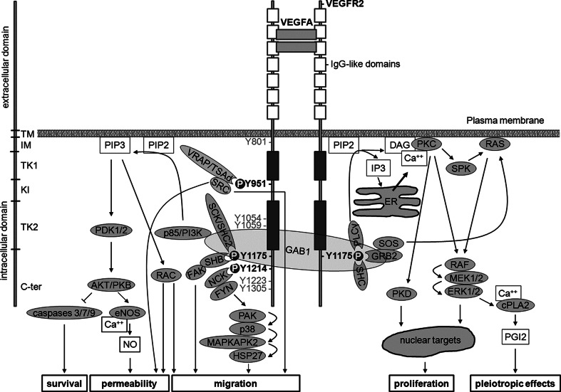
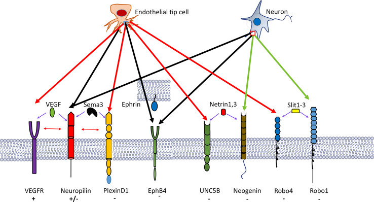
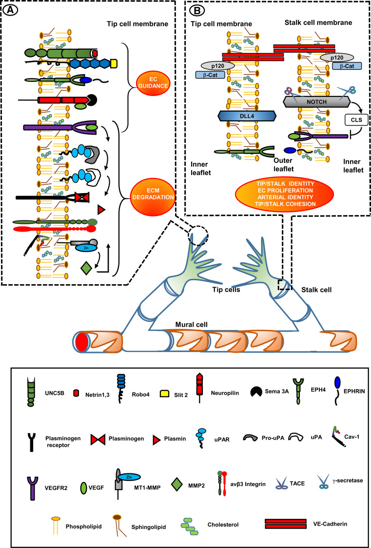
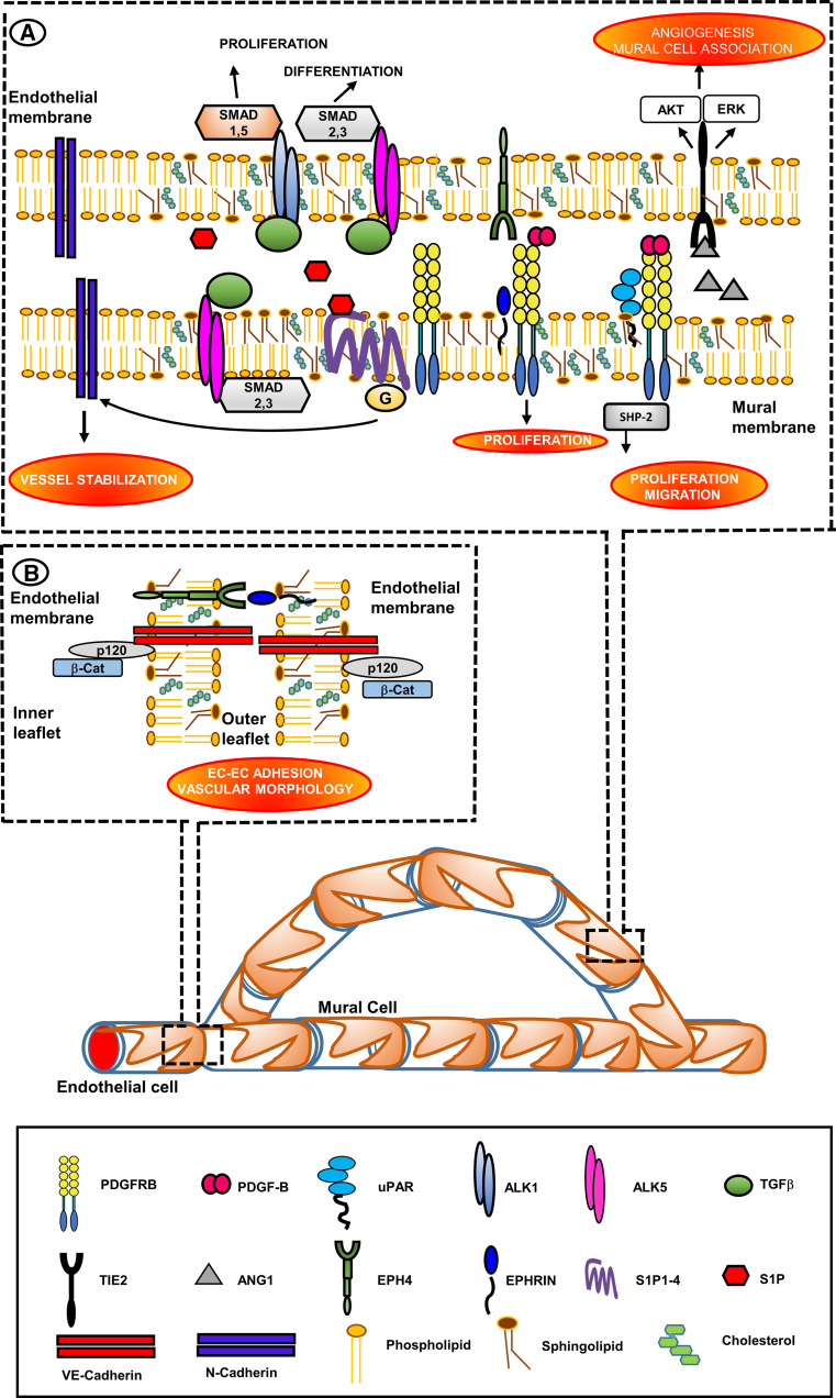
Research on the nanoscale membrane structures known as lipid rafts is relevant to the fields of cancer biology, inflammation and ischaemia. Lipid rafts recruit molecules critical to signalling and regulation of the invasion process in malignant cells, the leukocytes that provide immunity in inflammation and the endothelial cells that build blood and lymphatic vessels, as well as the patterning of neural networks. As angiogenesis is a common denominator, regulation of receptors and signalling molecules critical to angiogenesis is central to the design of new approaches aimed at reducing, promoting or normalizing the angiogenic process. The goal of this review is to highlight some of the key issues that indicate the involvement of endothelial cell lipid rafts at each step of so-called 'sprouting angiogenesis', from stimulation of the vascular endothelial growth factor to the choice of tip cells, activation of migratory and invasion pathways, recruitment of molecules that guide axons in vascular patterning and maturation of blood vessels. Finally, the review addresses opportunities for future studies to define how these lipid domains (and their constituents) may be manipulated to stimulate the so-called 'normalization' of vascular networks within tumors, and be identified as the main target, enabling the development of more efficient chemotherapeutics and cancer immunotherapies.


Figures

Fig. 1
Fig. 1
VEGF signalling elicited by VEGFR2-phosphorylating events. Structure and domains of VEGFR2 are indicated on the left side. Dimerized VEGFA (or VEGFC or -D after proteolytic cleavage) binds to second and third IgG-like domains within the extracellular domain of VEGFR2, thus inducing VEGFR2 dimerization and autophosphorylation at several tyrosine residues within the intracellular domain. Signalling elicited by major tyrosine residues phosphorylated in human VEGFR2 951, 1175 and 1214 (corresponding to 949, 1173 and 1212, respectively, in mice) is described. Moreover, phosphorylation at Y1054 and Y1059 is required to achieve maximal kinase activity. Additional tyrosine phosphorylation may occur at Y1223, Y1305, Y1309 and Y1319 residues. However, the role of these events is still unknown. Autophosphorylation at Y801 may precede phosphorylation of Y1054 and Y1059. SH2 domain-containing proteins (grooved oval) interact with tyrosine phosphorylated (P) residues thus activating signalling pathways that elicit several biological effects including increased permeability, survival, migration and proliferation (lower boxes). TM transmembrane, IM intramembrane, TK tyrosine kinase, KI kinase insert, C-term C-terminal, VEGFA vascular endothelial growth factor A, VEGFR2 VEGF receptor 2, VRAP VEGFR-associated protein also known as TSAd, T cell-specific adaptor, SRC sarcoma, GAB1 GRB2- (growth factor receptor-bound protein 2) associated binder 1, GRB2 growth factor receptor-bound protein 2, PLC-γ phospholipase C γ, RAC Ras-related C3 botulinum toxin substrate, PIP3 phosphatidylinositol 3,4,5-trisphosphate, PI3K phosphatidylinositol 3 kinase, PDK phosphoinositide-dependent kinase, PKB protein kinase B also known as AKT, RAC-alpha serine/threonine-protein kinase, BAD BCL2 associated death promoter, BCL2 B-cell CLL/lymphoma 2, eNOS endothelial nitric oxide synthase, ER endoplasmic reticulum, PLC-γ phospholipase C γ, NO nitric oxide, FAK focal adhesion kinase, SHC src homology/collagen, SCK SHC-like protein also known as SHC2, (Src homology 2 domain containing) transforming protein), PIP2 phosphatidylinositol (4,5)-bisphosphate, IP3 inositol (1,4,5)-trisphosphate, DAG sn-1,2-diacylglycerol, PKC protein kinase C, ERK1/2 extracellular regulated kinases 1 and 2, MEK MAPK/Erk kinase, HSP27 heat-shock protein 27, MAPKAP 2/3 MAPK-activating protein kinases 2 and 3, PAK p21-activated protein kinase, p38 p38 mitogen-activated protein kinase, Ca ++ calcium, cPLA2 cytosolic phospholipase A2, SHB SH2 domain-containing adapter protein B. SPK sphingosine kinase, CDC42 cell division cycle 42
Fig. 2
Fig. 2
A number of neural guidance molecular pathways have been recognized to participate in blood vessel branching morphogenesis. These pathways include semaphorin-3A (Sema3A)–plexinD1, ephrinB2–EphB4 and SLIT2–Robo4, which all elicit EC chemorepulsion, whereas SLIT2–Robo1 and VEGF–VEGFR2 (and possibly VEGF–NRP1) have been implicated in endothelial tip cell chemoattraction and elongation. Predominantly EC-expressed receptors are indicated by red arrows, receptors with shared expression in the nervous and the vascular system by black arrows, and molecules with uncertain expression in the vascular system by green arrows. Symbols plus and minus indicate chemoattraction and chemorepulsion, respectively
Fig. 3
Fig. 3
Vessel development: lipid-raft-localized molecules involved in vessel branching morphogenesis. Therapeutic targets. Insets show selected LRs of the tip-cell filopodia (inset a) and of the tip-cell/stalk-cell interface (inset b). Inset 1 VEGFR2 is located in filopodia [59, 60]. EC guidance receptors are in LRs and their cognate ligands generally induce EC repulsion [140, 144, 145, 147, 148]. VEGF family members also bind to a different extracellular sequence of Nrps, resulting in attraction of vascular structures [155] in a way similar to VEGFA-VEGFR2 interaction. Upon VEGFA/VEGFR2 interaction, MT1-MMP is associated with cav-1 and integrin αvβ3 in caveolar-LRs of filopodia, where it activates pro-MMP2 to MMP2 that is then bound to β3 integrins in LRs [95, 96, 126]. MMP2 activated uPAR-bound pro-uPA to uPA [124], which cleaves the proenzyme plasminogen to yield active plasmin [125] that in turn activates pro-MMPs to active MMPs, as well as pro-uPA to active uPA [–121]. Inset 2 the Notch system is located in LRs of the stalk cell. VEGFR2 stimulation upregulates overexpression of the Notch ligand protein Dll4 in the rear moiety of the tip-cell, which leads to CSL-mediated silencing of VEGFR2 expression in stalk-cell [71]. The stalk cell becomes insensitive to VEGF stimulation. Tip-cell/stalk-cell connection is warranted by VE-cadherins (not associated to LRs), and by the LR-associated Eph-Ephrin system [2]. The redorange ellipses identify the LR-directed processes, important in vessel development, that may be potentiated by forcing induction of LR formation to promote functional recovery and normalization of angiogenesis in tumors and other angiogenesis diseases
Fig. 4
Fig. 4
Vessel maturation: lipid-rafts as platforms of vessel stabilization by promotion of EC/mural cells and EC/EC interactions. Therapeutic targets. Insets show the LRs-located molecules that promote vessel stabilization and maturation by allowing mural cells (pericytes in capillaries and vSMS in larger vessels)/ECs (inset a) and EC/EC interactions (inset b). Inset a PDGFRβ is located in LRs of mural cells [181]. PDGFRβ requires the presence of partners for a proper signaling. uPAR associates with PDGFRβ in vSMCs. Assembly of this complex is necessary for signaling and initiation of functional changes in vSMCs mediated by the tyrosine phosphatase SHP-2. The complex is assembled on LRs [182, 183]. In vSMCs Ephrin-B2 interacts with PDGFRβ and controls its distribution and signaling, thereby promoting vSMCs proliferation [184]. PDGFRβ function also involves cooperation with LR-associated S1P receptors [185], thereby stimulating EC recruitment of pericytes and vSMCs [186]. S1P derives from EC sphingolipids that are marker constituents of LRs [187]. The main role of S1P1 receptor involves the trafficking of the cell adhesion molecule N-cadherin, which is not a LR-associated molecule, to the EC-mural-cell contact zone [188]. Tie2 associates with LRs following stimulation with Ang1 [193]. Caveolar-LRs facilitate the degradation of TGFβ receptors and therefore the turn-off of TGFβ signaling [190], thus controlling excess TGFβ activity. Inset b VE-cadherin is specific to EC adherent junctions and is necessary for vascular morphogenesis. VE-cadherin is associated to LRs. VE-cadherin is in turn associated with p120-catenin, and this interaction is necessary for VE-cadherin recruitment in LRs [13]. Ephrins and Eph receptors form clusters in LRs, providing low-affinity ephrin–ephrin and Eph–Eph dimers. Ephrin docking triggers stable aggregation into larger Eph-ephrin clusters which may fuse together into larger signaling platforms following Eph receptor-ephrin binding [, –173]. The red–orange ellipses identify the LR-directed processes that have the chance to favor EC–EC interaction (to prevent endothelial fenestrations and vascular leakage), and vessel wall integrity upon EC-mural cells interaction. Even if N-cadherins are not associated to LRs, their overexpression is a LR-dependent event directed by S1P1 receptor. Potentiation of LR formation may therefore have the chance to promote functional recovery and normalization of angiogenesis in tumors and other angiogenesis diseases where endothelial fenestrations, vascular leakage and loss of mural cells compromise vessel wall integrity

References

    1. Carmeliet P. Angiogenesis in health and disease. Nat Med. 2003;9:653–660. - PubMed
    1. Adams RH, Alitalo K. Molecular regulation of angiogenesis and lymphangiogenesis. Nat Rev Mol Cell Biol. 2007;8:464–478. - PubMed
    1. Roura S, Gálvez-Montón C, Bayes-Genis A. The challenges for cardiac vascular precursor cell therapy: lessons from a very elusive precursor. J Vasc Res. 2013;50:304–323. - PubMed
    1. Logsdon EA, Finley SD, Popel AS, et al. A systems biology view of blood vessel growth and remodelling. J Cell Mol Med. 2014 - PMC - PubMed
    1. Gerhardt H, Betsholtz C. How do endothelial cells orientate? Experientia. 2005;94:3–15. - PubMed

Publication types

Substances