. 2015 May;25(5):570-81.
doi: 10.1093/glycob/cwu188. Epub 2014 Dec 30.

The structures of glycophorin C N-glycans, a putative component of the GPC receptor site for Plasmodium falciparum EBA-140 ligand

Affiliations

The structures of glycophorin C N-glycans, a putative component of the GPC receptor site for Plasmodium falciparum EBA-140 ligand

David J Ashline et al. Glycobiology. 2015 May.

Abstract

Glycophorins C and D are highly glycosylated integral sialoglycoproteins of human red blood cell membranes carrying the Gerbich blood group antigens. The O- and N-glycosidic chains of the major erythrocyte glycoprotein (Lisowska E. 2001, Antigenic properties of human glycophorins - an update. Adv Exp Med Biol, 491:155-169; Tomita M and Marchesi VT. 1975, Amino-acid sequence and oligosaccharide attachment sites of human erythrocyte glycophorin. Proc Natl Acad Sci USA, 72:2964-2968.) are well characterized but the structure of GPC N-glycans has remained unknown. This problem became important since it was reported that GPC N-glycans play an essential role in the interaction with Plasmodium falciparum EBA-140 merozoite ligand. The elucidation of these structures seems essential for full characterization of the GPC binding site for the EBA-140 ligand. We have employed detailed structural analysis using sequential mass spectrometry to show that many GPC N-glycans contain H2 antigen structures and several contain polylactosamine structures capped with fucose. The results obtained indicate structural heterogeneity of the GPC N-glycans and show the existence of structural elements not found in glycophorin A N-glycans. Our results also open a possibility of new interpretation of the data concerning the binding of P. falciparum EBA-140 ligand to GPC. We hypothesize that preferable terminal fucosylation of N-glycosidic chains containing repeating lactosamine units of the GPC Gerbich variant could be an explanation for why the EBA-140 ligand does not react with GPC Gerbich and an indication that the EBA-140 interaction with GPC is distinctly dependent on the GPC N-glycan structure.

Keywords: EBA-140 ligand; Plasmodium; glycan; glycophorin; mass spectrometry.


Figures

Fig. 1.
Fig. 1.
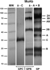
Western blotting of purified GPC+GPB sample used for N-glycan MS analysis detected with MoAb anti-GPC (clone NaM57-1F6) and anti-GPA+GPB (clone NaM26-3F4). The sample consists of GPC and GPB (which does not contain N-glycan) and GPA is not present. MW, protein molecular-weight standards; GPC+GPB sample; GP, crude glycophorins.
Fig. 2.
Fig. 2.
MALDI mass spectrum of reduced and permethylated N-linked glycans from GPC. Masses are labeled; structures and relative amounts are enumerated in Table I.
Fig. 3.
Fig. 3.
MSn spectra of the m/z 13512+, composition Hex5HexNAc5dHex2, shows a biantennary structure with one core fucose and one antennal fucose. (A) The MS2 spectrum of the doubly-charged m/z 1351. The structure is shown in (G). (B) The MS3 of the doubly-charged m/z 1221. (C) The MS4 of the doubly-charged m/z 990. This structure is shown in (H), reflecting the loss of bisecting GlcNAc and terminal LacNAc. Fragmentation of the core m/z 852 fragment in (D) and (E) is indicative of a structure having a bisecting GlcNAc as shown in (I). The fucosylated antenna fragment was isolated as the B-type ion, m/z 660, shown in (F). This fragmentation pattern is consistent with an H2 structure (J), with no trace of Lewis X. Normalization level (NL) and signal averaging time are shown for each spectrum.
Fig. 4.
Fig. 4.
MSn spectra of m/z 14442+, composition Hex5HexNAc5dHexNeuAc, showing a biantennary structure with two isomers, differing in fucose location. (A) The MS2 spectrum of the m/z 1444 precursor. (B) The MS3 (m/z 1444 → 1257) spectrum of the fragment formed by loss of NeuAc. (C) The MS4 (m/z 1444 → 1257 → 1127) spectrum of the fragment formed by successive losses of NeuAc and terminal HexNAc. These spectra show isomers with both core Fuc-GlcNAc (m/z 490) and antennal Fuc (m/z 660). (D) and (E) The core fragments indicating a structure containing a bisecting GlcNAc. (F) and (G) The putative structures of these two isomers. (H) The structure of the isolated H2 B-type ion with fragment assignments. (I) The MS3 m/z 660, a terminal fucosylated lactosamine ion, with fragments consistent with H2, based on similarity to standards and de novo fragment assignment.
Fig. 5.
Fig. 5.
Partial MSn data set for m/z 16632+, composition Hex6HexNAc6dHex3. Disassembly reveals at least two isomers, differing in antenna number and length, but both having bisecting GlcNAc. Differing antennal numbers are verified by the presence of both m/z 852 and 838 fragment ions, indicative of biantennary (with bisecting GlcNAc) and triantennary (with bisecting GlcNAc). As before, the m/z 660 ion and its CID spectrum are consistent with H2 epitope.
Fig. 6.
Fig. 6.
Disassembly of the extended antenna from Hex6HexNAc6dHex3, m/z 16632+. The singly charged fragment at m/z 1109 is consistent with a B-type monofucosylated diLacNAc structure, composition Hex2HexNAc2dHex1.

References

    1. Ashline D, Singh S, Hanneman A, Reinhold V. 2005. Congruent strategies for carbohydrate sequencing. 1. Mining structural details by MSn. Anal Chem. 77:6250–6262. - PMC - PubMed
    1. Ashline DJ, Hanneman AJ, Zhang H, Reinhold VN. 2014. Structural documentation of glycan epitopes: Sequential mass spectrometry and spectral matching. J Am Soc Mass Spectrom. 25:444–453. - PMC - PubMed
    1. Ashline DJ, Lapadula AJ, Liu YH, Lin M, Grace M, Pramanik B, Reinhold VN. 2007. Carbohydrate structural isomers analyzed by sequential mass spectrometry. Anal Chem. 79:3830–3842. - PMC - PubMed
    1. Ashline DJ, Yu Y, Lasanajak Y, Song X, Hu L, Ramani S, Prasad V, Estes MK, Cummings RD, Smith DF, et al. 2014. Structural characterization by multistage mass spectrometry (MSn) of human milk glycans recognized by human rotaviruses. Mol Cell Proteomics. 13:2961–2974. - PMC - PubMed
    1. Blanchard D, Dahr W, Hummel M, Latron F, Beyreuther K, Cartron JP. 1987. Glycophorins B and C from human erythrocyte membranes. Purification and sequence analysis. J Biol Chem. 262:5808–5811. - PubMed

Publication types