. 2015 Apr 15;24(8):2185-200.
doi: 10.1093/hmg/ddu738. Epub 2014 Dec 30.

Nephrocystin proteins NPHP5 and Cep290 regulate BBSome integrity, ciliary trafficking and cargo delivery

Affiliations

Nephrocystin proteins NPHP5 and Cep290 regulate BBSome integrity, ciliary trafficking and cargo delivery

Marine Barbelanne et al. Hum Mol Genet. .

Abstract

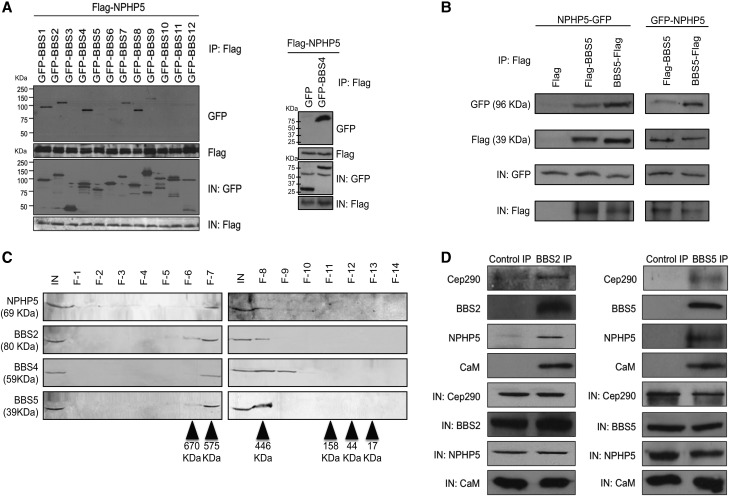
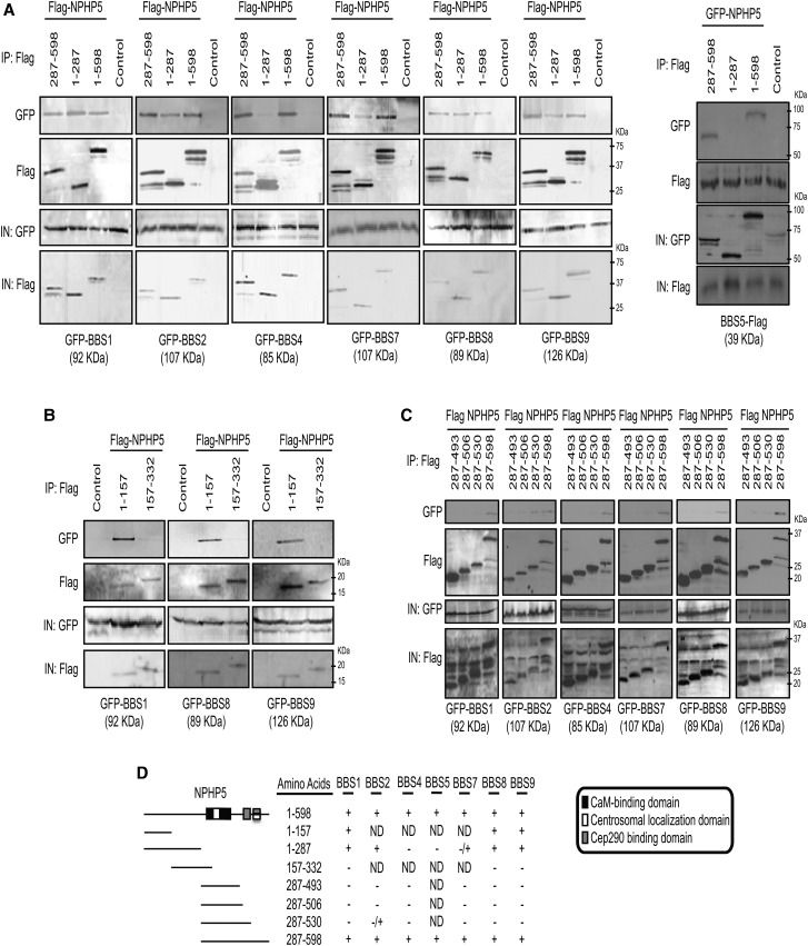
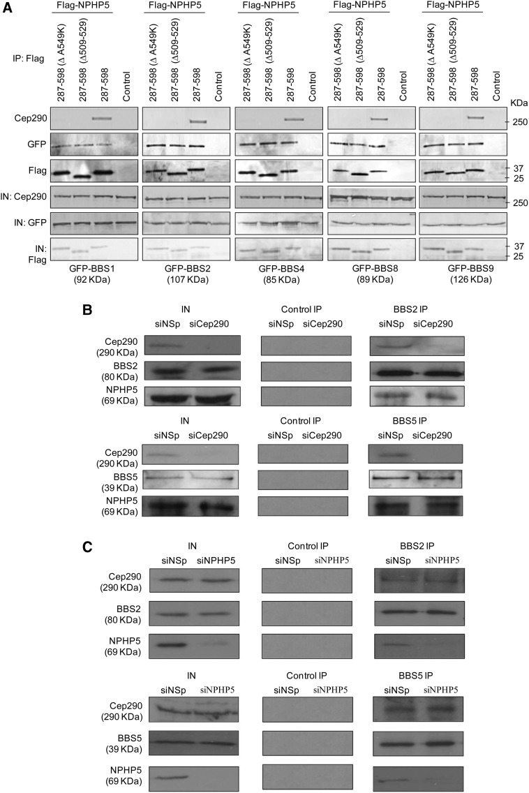
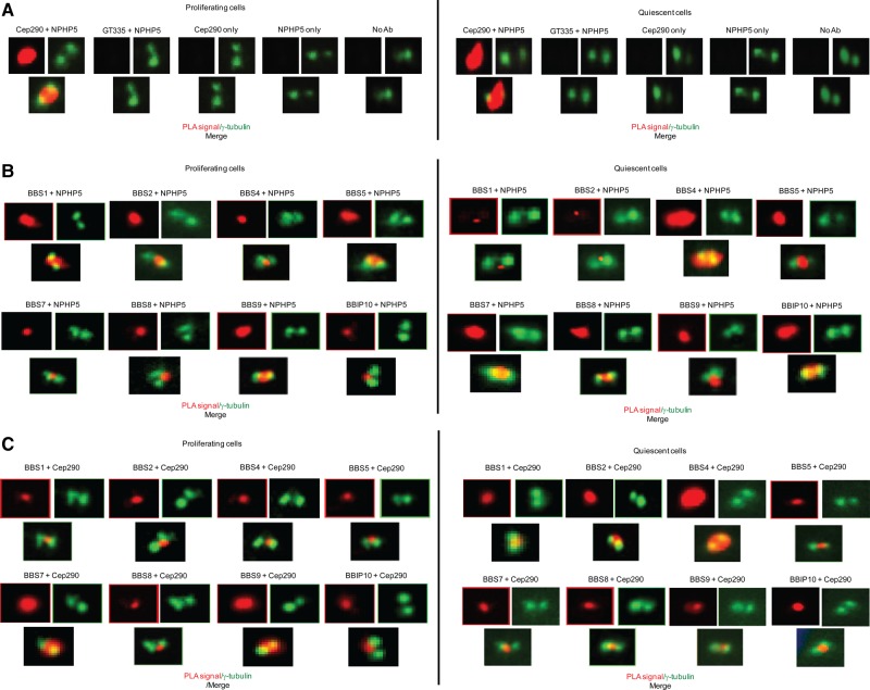
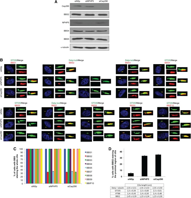
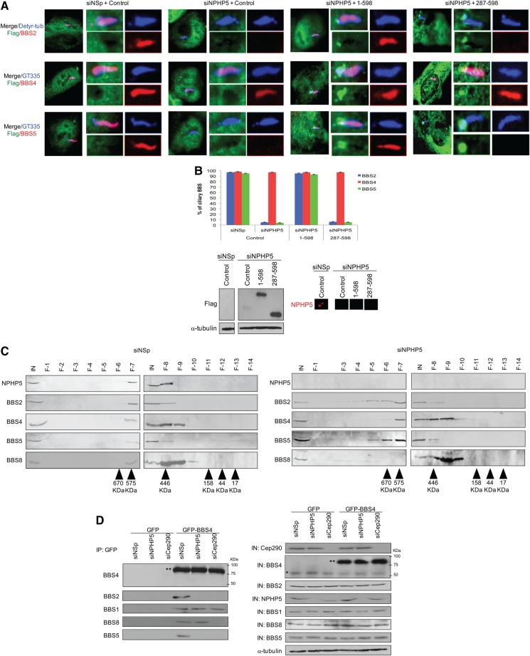
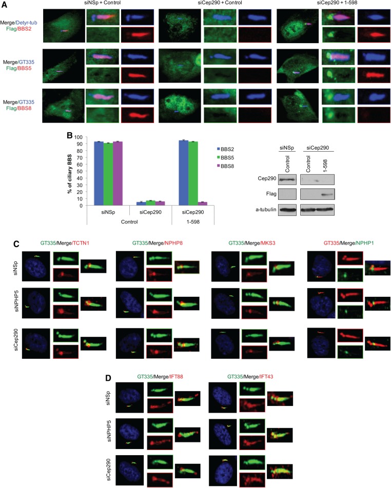
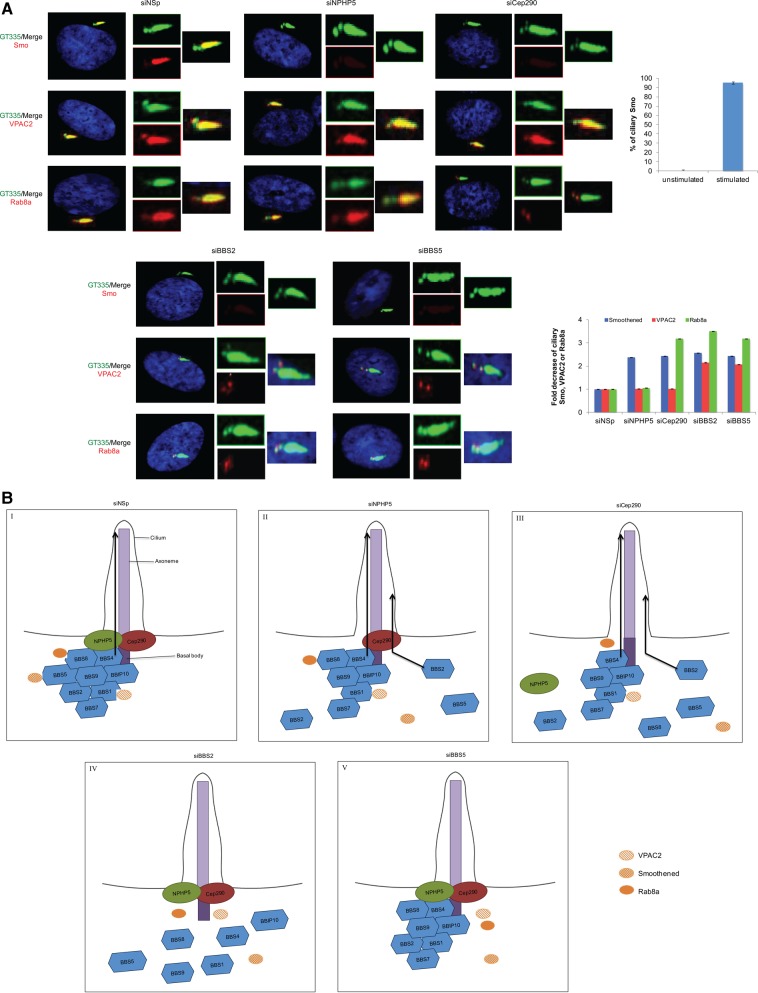
Proper functioning of cilia, hair-like structures responsible for sensation and locomotion, requires nephrocystin-5 (NPHP5) and a multi-subunit complex called the Bardet-Biedl syndrome (BBS)ome, but their precise relationship is not understood. The BBSome is involved in the trafficking of membrane cargos to cilia. While it is known that a loss of any single subunit prevents ciliary trafficking of the BBSome and its cargos, the mechanisms underlying ciliary entry of this complex are not well characterized. Here, we report that a transition zone protein NPHP5 contains two separate BBS-binding sites and interacts with the BBSome to mediate its integrity. Depletion of NPHP5, or expression of NPHP5 mutant missing one binding site, specifically leads to dissociation of BBS2 and BBS5 from the BBSome and loss of ciliary BBS2 and BBS5 without compromising the ability of the other subunits to traffic into cilia. Depletion of Cep290, another transition zone protein that directly binds to NPHP5, causes additional dissociation of BBS8 and loss of ciliary BBS8. Furthermore, delivery of BBSome cargos, smoothened, VPAC2 and Rab8a, to the ciliary compartment is completely disabled in the absence of single BBS subunits, but is selectively impaired in the absence of NPHP5 or Cep290. These findings define a new role of NPHP5 and Cep290 in controlling integrity and ciliary trafficking of the BBSome, which in turn impinge on the delivery of ciliary cargo.


Figures

Figure 1.
Figure 1.
NPHP5 interacts with the BBSome. (A) Flag-NPHP5 and the indicated GFP proteins were co-expressed in HEK293 cells, and lysates were immunoprecipitated with an anti-Flag antibody. The resulting immunoprecipitates were western blotted with anti-Flag or anti-GFP antibodies. IN, input. (B) The indicated N-terminal or C-terminal tagged recombinant NPHP5 or BBS5 proteins were co-expressed in HEK293 cells, and lysates were immunoprecipitated with an anti-Flag antibody. The resulting immunoprecipitates were western blotted with anti-Flag or anti-GFP antibodies. IN, input. (C) HEK293 cell extract was chromatographed on a Superose-6 gel filtration column, and the resulting fractions were western blotted with indicated antibodies. Estimated molecular weights are indicated. IN, input; F, fraction. (D) Western blotting of endogenous Cep290, NPHP5, BBS2, BBS5 and CaM after immunoprecipitation of HEK293 cell extracts with anti-Flag (control), anti-BBS2 or anti-BBS5 antibodies. IN, input.
Figure 2.
Figure 2.
NPHP5 possesses two distinct BBSome-binding sites. (A) Left panel: Flag (control), Flag-tagged full-length NPHP5 (1–598) or the indicated fragments of Flag-tagged NPHP5 were co-expressed with the indicated GFP-BBS proteins in HEK293 cells, and lysates were immunoprecipitated with an anti-Flag antibody. Flag-NPHP5 and GFP-BBS were detected after western blotting the resulting immunoprecipitates. IN, input. Right panel: GFP (control), GFP-tagged full-length NPHP5 (1–598) or the indicated fragments of GFP-tagged NPHP5 were co-expressed with BBS5-Flag in HEK293 cells, and lysates were immunoprecipitated with an anti-Flag antibody. BBS5-Flag and GFP-NPHP5 were detected after western blotting the resulting immunoprecipitates. IN, input. (B and C) Flag (control) or the indicated fragments of Flag-tagged NPHP5 were co-expressed with the indicated GFP-BBS proteins in HEK293 cells, and lysates were immunoprecipitated with an anti-Flag antibody. Flag-NPHP5 and GFP-BBS were detected after western blotting the resulting immunoprecipitates. IN, input. (D) Summary of the results of in vivo binding experiments. Known domains of NPHP5 (centrosomal localization, CaM-binding and Cep290-binding domains) are indicated as in (25). Each fragment used for the study takes into account the position of these domains so that a given domain is not prematurely truncated. +, interaction; –, no interaction; ND, not determined.
Figure 3.
Figure 3.
NPHP5 and Cep290 bind to the BBSome independently of each other. (A) Flag (control) or the indicated fragments and mutants of Flag-tagged NPHP5 were co-expressed with the indicated GFP-BBS proteins in HEK293 cells, and lysates were immunoprecipitated with an anti-Flag antibody. Flag-NPHP5, GFP-BBS and endogenous Cep290 were detected after western blotting the resulting immunoprecipitates. IN, input. (B and C) Western blotting of endogenous Cep290, NPHP5, BBS2 and BBS5 after immunoprecipitation of HEK293 cell extracts with anti-Flag (control), anti-BBS2 or anti-BBS5 antibodies. Extracts were transfected with control (siNSp), NPHP5 (siNPHP5) or Cep290 (siCep290) siRNAs prior to immunoprecipitation. IN, input.
Figure 4.
Figure 4.
NPHP5/Cep290 interaction with the BBSome in ciliated and non-ciliated cells. (A) In situ PLAs using antibodies against NPHP5 and Cep290 were performed to visualize protein–protein interaction (PLA signal in red) in proliferating (non-ciliated) and quiescent (ciliated) RPE-1 cells. Cells were co-stained with γ-tubulin (green) to visualize the centrosome. No PLA signal was detected when one or both antibodies were missing, or when an NPHP5 antibody was combined with an irrelevant antibody, glutamylated tubulin (GT335). No Ab, no antibody. (B and C) In situ PLAs using the indicated combinations of antibodies were performed to visualize protein–protein interaction (PLA signal in red) in proliferating (non-ciliated) and quiescent (ciliated) RPE-1 cells. Cells were co-stained with γ-tubulin (green) to visualize the centrosome.
Figure 5.
Figure 5.
Depletion of NPHP5 or Cep290 impairs ciliary localization of a subset of BBSome subunits. (A) Western blotting of Cep290, BBS2, BBS4, BBS5 and NPHP5 in HEK293 cells treated with control (siNSp), NPHP5 (siNPHP5) or Cep290 (siCep290) siRNAs. α-tubulin was used as loading control. (B) RPE-1 cells transfected with control (siNSp), NPHP5 (siNPHP5) or Cep290 (siCep290) siRNAs and induced to quiescence were stained with the indicated combinations of antibodies. Detyr-tub, detyrosinated tubulin; GT335, polyglutamylated tubulin. (C) The percentages of quiescent RPE-1 cells showing BBS staining along the entire length of cilia were determined using detyrosinated tubulin or GT335 as a ciliary marker. (D) (Top) The percentages of quiescent RPE-1 cells showing BBS2 staining along the proximal region of cilia were determined. (Bottom) Cilia length was measured with different markers (BBS2, detyrosinated tubulin, polyglutamylated tubulin and IFT88). In (C and D), at least 100 cells were counted and/or measured per siRNA condition, and error bars represent average of three independent experiments.
Figure 6.
Figure 6.
NPHP5 and Cep290 regulate BBSome integrity. (A) ARPE-19 cells transfected with control siRNA (siNSp) or siRNA targeting NPHP5 3′UTR (siNPHP5) and plasmid expressing an irrelevant Flag-tagged protein (control), full-length Flag-NPHP5 (1-598) or a C-terminal fragment of Flag-tagged NPHP5 (287-598) lacking a BBSome-binding site, induced to quiescence, were stained with antibodies against Flag (green), detyrosinated-tubulin or GT335 (blue), and BBS2, BBS4 or BBS5 (red). (B) (Top) The percentages of quiescent cells with ciliary BBS2, BBS4 or BBS5 staining were determined using either detyrosinated tubulin or GT335 as a ciliary marker. At least 100 transfected cells were counted per condition, and error bars represent average of three independent experiments. (Bottom left) Western blotting of Flag, with α-tubulin used as loading control. (Bottom right) Cells were processed for immunofluorescence and stained with anti-NPHP5 (red) antibody. (C) Extract from control (siNSp) or NPHP5 siRNA-depleted (siNPHP5) HEK293 cells was chromatographed on a Superose-6 gel filtration column, and the resulting fractions were western blotted with indicated antibodies. Estimated molecular weights are indicated. IN, input; F, fraction. (D) GFP or GFP-BBS4 were expressed in HEK293 cells treated with control (siNSp), NPHP5 (siNPHP5) or Cep290 (siCep290) siRNAs and immunoprecipitated from lysates. Endogenous (*) and recombinant BBS4 (**) along with endogenous BBS1, BBS2, BBS5 and BBS8 were detected after western blotting the resulting immunoprecipitates. Western blotting of NPHP5 and Cep290 were performed to monitor knockdown efficiency. IN, input. α-tubulin was used as loading control.
Figure 7.
Figure 7.
Cep290 regulates BBSome integrity and depletion of NPHP5 or Cep290 does not impair the localization of transition zone proteins. (A) ARPE-19 cells transfected with control (siNSp) or Cep290 (siCep290) siRNA and plasmid expressing an irrelevant Flag-tagged protein (control) or full-length Flag-NPHP5 (1-598), induced to quiescence, were stained with antibodies against Flag (green), detyrosinated-tubulin or GT335 (blue), and BBS2, BBS5 or BBS8 (red). (B) (Left) The percentages of quiescent cells with ciliary BBS2, BBS5 or BBS8 staining were determined using either detyrosinated tubulin or GT335 as a ciliary marker. At least 100 transfected cells were counted per condition, and error bars represent average of three independent experiments. (Right) Western blotting of Flag and Cep290. α-tubulin was used as loading control. (C and D) RPE-1 cells transfected with control (NSp), NPHP5 (siNPHP5) or Cep290 (siCep290) siRNAs and induced to quiescence were stained with the indicated antibodies.
Figure 8.
Figure 8.
Depletion of NPHP5 or Cep290 partially disrupts ciliary trafficking of BBSome cargos. (A) (Left) RPE-1 cells transfected with control (NSp), NPHP5 (siNPHP5), Cep290 (siCep290), BBS2 (siBBS2) or BBS5 (siBBS5) siRNAs, induced to quiescence, were stained with antibodies against Smo, VPAC2 or Rab8a (red) and GT335 (green). In the case of Smo, cells were treated with a Smo agonist prior to immunofluorescence. (Top right) The percentage of quiescent cells with ciliary Smo, untreated (unstimulated) or treated with a Smo agonist (stimulated), was determined using GT335 as a ciliary marker. (Bottom right) The fold decrease in the percentage of cells with ciliary Smo, VPAC2 or Rab8a was determined using GT335 as a ciliary marker. At least 100 cells were scored per condition, and error bars represent average of three independent experiments. (B) Model illustrating the roles of NPHP5 and Cep290 in BBSome homeostasis. (I) NPHP5 and Cep290 at the ciliary base serve two purposes: they regulate BBSome integrity and form a diffusion barrier that allows the selective passage of the holo-complex into the cilium. Although NPHP5 and Cep290 interact with multiple subunits, their interaction with BBS4/BBS8 is shown for the sake of simplicity. (II) In the absence of NPHP5, BBS2 and BBS5 dissociate from the BBSome, and BBS5 is completely mislocalized from the cilium. A fraction of BBS2 exhibits similar mislocalization pattern, whereas another fraction is confined to the proximal region of the cilium through an undefined mechanism. A faulty barrier permits the BBSome sub-complex to traffic into the cilium. Ciliary trafficking of Smo is impaired, since this cargo normally interacts with BBS5. (III) A loss of Cep290 induces NPHP5 mislocalization and promotes further dissociation of BBS8 from the BBSome; yet this sub-complex is also allowed to undergo ciliary trafficking. Rab8a ciliary trafficking is additionally impaired, but its centrosomal localization is unaffected. (IV and V) In contrast, a loss of any single BBS subunit, irrespective of its effects on BBSome assembly, is excluded from the cilium due to an intact barrier created by NPHP5/Cep290. Inactivation of BBS2 is known to destabilize BBS7 and prevents early BBSome assembly, whereas inactivation of BBS5 has minor impact on assembly (22,59). As a consequence, Smo, Rab8a and VPAC2 are unable to enter the ciliary compartment, and Rab8a and VPAC2 remain at the centrosome. We speculate that Rab8a is transported into the cilium by tethering to BBS8, whereas ciliary trafficking of VPAC2 likely requires interaction with BBS1, BBS4, BBS7, BBS9 and/or BBIP10. The interaction between VPAC2 and BBS1 is shown for simplicity.

Similar articles

Cited by

References

    1. Bornens M. (2012) The centrosome in cells and organisms. Science, 335, 422–426. - PubMed
    1. Hossain D., Tsang W.Y. (2013) Centrosome dysfunction and senescence: coincidence or causality? Aging Sci., 1, 113.
    1. Nigg E.A., Stearns T. (2011) The centrosome cycle: centriole biogenesis, duplication and inherent asymmetries. Nat. Cell Biol., 13, 1154–1160. - PMC - PubMed
    1. Debec A., Sullivan W., Bettencourt-Dias M. (2010) Centrioles: active players or passengers during mitosis? Cell Mol. Life Sci., 67, 2173–2194. - PMC - PubMed
    1. Kim S., Dynlacht B.D. (2013) Assembling a primary cilium. Curr. Opin. Cell Biol., 25, 506–511. - PMC - PubMed

Publication types

MeSH terms