. 2015 Mar 15;308(6):R507-16.
doi: 10.1152/ajpregu.00406.2014. Epub 2014 Dec 31.

Genetic knockdown of estrogen receptor-alpha in the subfornical organ augments ANG II-induced hypertension in female mice

Affiliations

Genetic knockdown of estrogen receptor-alpha in the subfornical organ augments ANG II-induced hypertension in female mice

Baojian Xue et al. Am J Physiol Regul Integr Comp Physiol. .

Abstract

The present study tested the hypotheses that 1) ERα in the brain plays a key role in the estrogen-protective effects against ANG II-induced hypertension, and 2) that the subfornical organ (SFO) is a key site where ERα mediates these protective actions. In this study, a "floxed" ERα transgenic mouse line (ERα(flox)) was used to create models in which ERα was knocked down in the brain or just in the SFO. Female mice with ERα ablated in the nervous system (Nestin-ERα(-) mice) showed greater increases in blood pressure (BP) in response to ANG II. Furthermore, females with ERα knockdown specifically in the SFO [SFO adenovirus-Cre (Ad-Cre) injected ERα(flox) mice] also showed an enhanced pressor response to ANG II. Immunohistochemical (IHC), RT-PCR, and Western blot analyses revealed a marked reduction in the expression of ERα in nervous tissues and, in particular, in the SFO. These changes were not present in peripheral tissues in Nestin-ERα(-) mice or Ad-Cre-injected ERα(flox) mice. mRNA expression of components of the renin-angiotensin system in the lamina terminalis were upregulated in Nestin-ERα(-) mice. Moreover, ganglionic blockade on day 7 after ANG II infusions resulted in a greater reduction of BP in Nestin-ERα(-) mice or SFO Ad-Cre-injected mice, suggesting that knockdown of ERα in the nervous system or the SFO alone augments central ANG II-induced increase in sympathetic tone. The results indicate that interfering with the action of estrogen on SFO ERα is sufficient to abolish the protective effects of estrogen against ANG II-induced hypertension.

Keywords: ANG II; blood pressure; estrogen receptor-α; nervous system; subfornical organ.


Figures

Fig. 1.
Fig. 1.
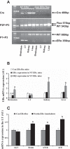
Representative tissue genotyping and mRNA expression of ERα, ERβ, and renin-angiotensin system (RAS) components in Nestin-ERα and Con-ERαflox mice. A, top: Cre is present in all of the tissues of Nestin-ERα mouse, but is absent in Con-ERαflox mouse. Middle: shown are flox band (575 bp) and ERα wild-type (WT) band by using a combination of ERα P2F and P3 primers, which detects the presence of LoxP sequences. All tissues had WT bands (543 bp), whereas flox bands (575 bp) were removed only in the brain stem and lamina terminalis (LT) in the Nestin-ERαmouse. Bottom: bands detected by combination of ERα P1 and P3 primers, which confirms whether exon 3 of Nestin-ERα mouse was deleted in the presence of Cre recombinase. As expected, a smaller DNA band (359 bp) was present in the brain stem and LT of Nestin-ERα mouse with the presence of Cre. This did not occur in kidney and liver of Nestin-ERα mouse and any checked tissues of the Con-ERαflox mouse. B: mRNA expression of ERα in the brain stem and LT, but not in kidney and liver, was significantly knocked down in Nestin-ERα mice, while ERβ expression in the brain stem, kidney, and liver, but not in the LT, was significantly upregulated compared with that in Con-ERαflox mice. C: upregulation of mRNA expression in RAS components in the LT of Nestin-ERα when compared with Con-ERαflox mice. (*P < 0.05 vs. Con-ERαflox mice).
Fig. 2.
Fig. 2.
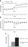
The nervous tissue-specific knockdown of ERα augmented the pressor effect of ANG II treatment. Daily measurement of mean arterial pressure (MAP; A) and heart rate (HR; B) before and during infusion of ANG II in Con-ERαflox and Nestin-ERα mice. C: averaged increases in MAP induced by ANG II infusion in these two groups of mice. Control days are denoted by the letter C, followed by 7 days of ANG II infusion. *P < 0.05 compared with baseline. #P < 0.05 compared with Con-ERαflox mice given ANG II.
Fig. 3.
Fig. 3.
SFO ERα-mediated the protective effect of estrogen in ANG II-induced hypertension. Daily measurements of MAP (A) and HR (B) before and during infusion of ANG II in SFO Ad-Cre or Ad-Con treated ERαflox mice. C: average changes in MAP across days induced by ANG II infusion. Control days are denoted by the letter C, followed by 7 days of ANG II infusion. *P < 0.05 vs. baseline. #P < 0.05 vs. Ad-Con group.
Fig. 4.
Fig. 4.
A: photomicrograph that illustrates the anatomical placement of the subfornical organ (SFO) cannula and the injection site. SFO Ad-Cre-injected ERαflox mice showed reduced ERα staining (green) in the SFO that paralleled intense Cre staining (red) in this site. The injections were targeted to the ventral hippocampal commissure so that injectate would diffuse into the SFO to minimize mechanical damage to the SFO proper (B). C: robust ERα expression was seen in the OVLT or AP/NTS, but Cre expression could not be detected in these sites, indicating that the injected Ad-Cre was not likely to have leaked into the brain ventricle.
Fig. 5.
Fig. 5.
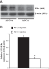
Western blot analysis of ERα protein expression in the SFO from mice receiving SFO injection of Ad-Cre or Ad-Con. A: representative Western blots of ERα and β-actin are shown. B: results of Western blot analysis represented the change in ERα protein expression, which was normalized with β-actin in the SFO.
Fig. 6.
Fig. 6.
Decreases in MAP in response to ganglionic blockade with hexamethonium before and on day 7 after infusion of ANG II in all groups of female mice. *P < 0.05 compared with control. #Compared to Con-ERαflox or Ad-Cre-injected ERαflox mice given ANG II.

Similar articles

Cited by

References

    1. Antonson P, Omoto Y, Humire P, Gustafsson JÅ. Generation of ERα-floxed and knockout mice using the Cre/LoxP system. Biochem Biophys Res Commun 424: 710–716, 2012. - PubMed
    1. Caldon CE. Estrogen signaling and the DNA damage response in hormone-dependent breast cancers. Front Oncol 4: 106, 2014. - PMC - PubMed
    1. Ciriello J, Roder S. 17β-Estradiol alters the response of subfornical organ neurons that project to supraoptic nucleus to plasma angiotensin II and hypernatremia. Brain Res 1526: 54–64, 2013. - PubMed
    1. Dean SA, Tan J, O'Brien ER, Leenen FH. 17 β-estradiol downregulates tissue angiotensin-converting enzyme and ANG II type 1 receptor in female rats. Am J Physiol Regul Integr Comp Physiol 288: R759–R766, 2005. - PubMed
    1. Dubois NC, Hofmann D, Kaloulis K, Bishop JM, Trumpp A. Nestin-Cre transgenic mouse line Nes-Cre1 mediates highly efficient Cre/LoxP mediated recombination in the nervous system, kidney, and somite-derived tissues. Genesis 44: 355–360, 2006. - PubMed

Publication types

MeSH terms