. 2015 Mar;89(6):3026-37.
doi: 10.1128/JVI.03432-14. Epub 2014 Dec 31.

Reverse genetics system for severe fever with thrombocytopenia syndrome virus

Affiliations

Reverse genetics system for severe fever with thrombocytopenia syndrome virus

Benjamin Brennan et al. J Virol. 2015 Mar.

Abstract

Severe fever with thrombocytopenia syndrome virus (SFTSV) is an emerging tick-borne pathogen that was first reported in China in 2009. Phylogenetic analysis of the viral genome showed that SFTS virus represents a new lineage within the Phlebovirus genus, distinct from the existing sandfly fever and Uukuniemi virus groups, in the family Bunyaviridae. SFTS disease is characterized by gastrointestinal symptoms, chills, joint pain, myalgia, thrombocytopenia, leukocytopenia, and some hemorrhagic manifestations with a case fatality rate of about 2 to 15%. Here we report the development of reverse genetics systems to study STFSV replication and pathogenesis. We developed and optimized functional T7 polymerase-based M- and S-segment minigenome assays, which revealed errors in the published terminal sequences of the S segment of the Hubei 29 strain of SFTSV. We then generated recombinant viruses from cloned cDNAs prepared to the antigenomic RNAs both of the minimally passaged virus (HB29) and of a cell culture-adapted strain designated HB29pp. The growth properties, pattern of viral protein synthesis, and subcellular localization of viral N and NSs proteins of wild-type HB29pp (wtHB29pp) and recombinant HB29pp viruses were indistinguishable. We also show that the viruses fail to shut off host cell polypeptide production. The robust reverse genetics system described will be a valuable tool for the design of therapeutics and the development of killed and attenuated vaccines against this important emerging pathogen.

Importance: SFTSV and related tick-borne phleboviruses such as Heartland virus are emerging viruses shown to cause severe disease in humans in the Far East and the United States, respectively. Study of these novel pathogens would be facilitated by technology to manipulate these viruses in a laboratory setting using reverse genetics. Here, we report the generation of infectious SFTSV from cDNA clones and demonstrate that the behavior of recombinant viruses is similar to that of the wild type. This advance will allow for further dissection of the roles of each of the viral proteins in the context of virus infection, as well as help in the development of antiviral drugs and protective vaccines.


Figures

FIG 1
FIG 1
Creation of HB29 S- and M-based minigenome constructs. (A) Schematic of the genome organization of S- and M-based minigenome constructs and the relative orientation of the open reading frames in the transcription plasmids pTVT7. (B) Effect of increasing amounts of pTM1-HB29N on M-segment minigenome assay. BSR-T7/5 cells were transfected with pTVT7-HB29M:hRen, pTM1-HB29ppL, pTM1-FF-Luc, and the indicated amount of pTM1-HB29N. (C) Effect of increasing amounts of pTM1-HB29ppL on M-segment minigenome assay. BSR-T7/5 cells were transfected with pTVT7-HB29M:hRen, pTM1-HB29N, pTM1-FF-Luc, and the indicated amount of pTM1-HB29ppL. In both cases, empty pTM1 vector was used to ensure that the total amount of DNA used in each transfection was the same (2.51 μg). Renilla luciferase activity was measured 24 h posttransfection.
FIG 2
FIG 2
Correction of the sequence of the S-segment-based minigenome. (A) Schematic representation of 3′ RACE data indicating that there is a missing A residue at position 9 of both the 5′ and 3′ S segment UTRs. Conserved terminal nucleotides are highlighted in red, and open reading frames for N and NSs proteins are underlined. (B) Effect of correcting the S-segment UTR sequence on S minigenome activity. BSR-T7/5 cells were transfected with pTM1-HB29N, pTM1-HB29ppL, pTM1-FF-Luc and contained either the published UTR sequences (Pub-UTR) or pTVT7-HB29SdelNSs:hRen with an A residue inserted at position 9 of the 5′ antigenomic UTR (5′AG UTR) or pTVT7-HB29SdelNSs:hRen with A and T residues inserted at position 9 of both 5′ and 3′ UTRs (Both UTR). “No L” is a negative control without pTM1-HB29ppL (empty pTM1 vector was used instead). Renilla luciferase activity was measured 24 h posttransfection. (C to F) eGFP autofluorescence observed in cells 24 h after transfection with analogous minigenome plasmids containing eGFP in place of hRen sequences: HB29 S with published UTR sequences (C); A residue inserted at position 9 of the 5′ antigenomic UTR (D); A and T residues inserted at position 9 of both 5′ and 3′ UTRs (E); negative control without pTM1-HB29ppL (F).
FIG 3
FIG 3
Effect of HB29 NSs protein on minigenome activity. BSR-T7/5 cells were transfected with pTM1-HB29ppL, pTM1-HB29N, pTM1-FF-Luc, the indicated increasing amounts of pTM1-HB29NSs, and either pTVT7-HB29SdelNSs:hRen (A) or pTVT7-HB29M:hRen (B). No L, negative control without pTM1-HB29ppL; empty pTM1 vector was used to ensure that equal amounts of DNA were transfected into each reaction mixture. Luciferase activities were measured at 24 h posttransfection.
FIG 4
FIG 4
Comparison of immunostained foci and plaque sizes of recombinant viruses. Vero E6 cells were infected with serial dilutions of virus and incubated under an 0.6% Avicel overlay for 7 days at 37°C. Cell monolayers were fixed with 4% formaldehyde. Following fixation, cell monolayers were subjected to immunofocus-staining assay or stained with Giemsa stain to visualize plaques. (A) Foci from rescue of cDNA clones representing minimally passaged HB29. (B, C) Increase in the number of large foci following passage of recombinant virus. (D, E) Direct comparison of rescue of recombinant virus from cDNA from minimally passaged HB29 (D) and from M segment cDNA containing the F330S mutation in Gn (E). (F to I) Direct comparison of immunostained foci formed by recombinant rHB29pp, authentic wtHB29pp, rHB29M:F330S, and rHB29 viruses, as indicated. (J to M) Direct comparison of Giemsa-stained plaques formed by recombinant rHB29pp, authentic wtHB29pp, rHB29M:F330S, and rHB29, as indicated.
FIG 5
FIG 5
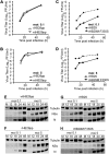
Growth properties of recombinant viruses. Viral growth curves were determined for Vero E6 cells infected at low (0.1) or high (5) MOI, and titers were measured by plaque assay or immunofocus assay as appropriate. (A, B) Comparison of recombinant and wild-type HB29pp. (C, D) Comparison of recombinant rHB20 and rHB29M:F330S. Graphs show a representative experiment of titrations carried out at the same time. (E to H) Western blot analysis of S-segment-encoded proteins from infected cells. Cell extracts were prepared from the growth curve samples at the time points indicated, proteins were fractionated on 4 to 12% NuPage gels, and blots were probed with anti-N, anti-NSs, and antitubulin antibodies as indicated.
FIG 6
FIG 6
Lack of inhibition of host cell protein synthesis. Vero E6 cells were infected at an MOI of 5 with wtHB29pp, rHB29pp, rHB29, or rHB29M:F330S or were mock infected (M) as indicated. Cells were labeled with 30 μCi [35S]methionine and/or cysteine for 2 h at the time points indicated, and cell extracts were fractionated by SDS-PAGE. The positions of the viral N (black arrows) and NSs (asterisks) proteins are shown.
FIG 7
FIG 7
Intracellular localization of N and NSs proteins in infected cells. Vero E6 cells were infected at an MOI of 5 with rHB29, wtHB29pp, rHB29pp, or rHB29M:F330S or mock infected. At 24 h p.i., the cells were fixed with 4% formaldehyde, followed by staining with either anti-N (A) or anti-NSs (B) monospecific antibodies. Samples were counterstained with DAPI. Cells were examined with a Zeiss LSM 710 confocal microscope. Bar, 10 μm.

References

    1. Plyusnin A, Beaty BJ, Elliott RM, Goldbach R, Kormelink R, Lundkvist Å, Schmaljohn CS, Tesh RB. 2012. Bunyaviridae, p 725–741 InKing AMQ, Adams MJ, Carstens EB, Lefkowits EJ (ed), Virus taxonomy: ninth report of the International Committee on Taxonomy of Viruses. Elsevier Academic Press, London, United Kingdom.
    1. Plyusnin A, Elliott RM (ed). 2011. Bunyaviridae: molecular and cellular biology. Caister Academic Press, Norfolk, United Kingdom.
    1. Walter CT, Barr JN. 2011. Recent advances in the molecular and cellular biology of bunyaviruses. J Gen Virol 92:2467–2484. doi:10.1099/vir.0.035105-0. - DOI - PubMed
    1. Elliott RM, Schmaljohn CS. 2013. Bunyaviridae, p 1244–1282 InKnipe DM, Howley PM (ed), Fields virology, 6th ed Wolters Kluwer, Philadelphia, PA.
    1. Bouloy M. 2011. Molecular biology of phleboviruses, p 95–128 InPlyusnin A, Elliott RM (ed), Bunyaviridae: molecular and cellular biology. Caister Academic Press, Norfolk, United Kingdom.

Publication types

Associated data