. 2015 Mar;89(6):3163-75.
doi: 10.1128/JVI.03353-14. Epub 2014 Dec 31.

Differential effects of the G118R, H51Y, and E138K resistance substitutions in different subtypes of HIV integrase

Affiliations

Differential effects of the G118R, H51Y, and E138K resistance substitutions in different subtypes of HIV integrase

Peter K Quashie et al. J Virol. 2015 Mar.

Abstract

Dolutegravir (DTG) is the latest antiretroviral (ARV) approved for the treatment of human immunodeficiency virus (HIV) infection. The G118R substitution, previously identified with MK-2048 and raltegravir, may represent the initial substitution in a dolutegravir resistance pathway. We have found that subtype C integrase proteins have a low enzymatic cost associated with the G118R substitution, mostly at the strand transfer step of integration, compared to either subtype B or recombinant CRF02_AG proteins. Subtype B and circulating recombinant form AG (CRF02_AG) clonal viruses encoding G118R-bearing integrases were severely restricted in their viral replication capacity, and G118R/E138K-bearing viruses had various levels of resistance to dolutegravir, raltegravir, and elvitegravir. In cell-free experiments, the impacts of the H51Y and E138K substitutions on resistance and enzyme efficiency, when present with G118R, were highly dependent on viral subtype. Sequence alignment and homology modeling showed that the subtype-specific effects of these mutations were likely due to differential amino acid residue networks in the different integrase proteins, caused by polymorphic residues, which significantly affect native protein activity, structure, or function and are important for drug-mediated inhibition of enzyme activity. This preemptive study will aid in the interpretation of resistance patterns in dolutegravir-treated patients.

Importance: Recognized drug resistance mutations have never been reported for naive patients treated with dolutegravir. Additionally, in integrase inhibitor-experienced patients, only R263K and other previously known integrase resistance substitutions have been reported. Here we suggest that alternate resistance pathways may develop in non-B HIV-1 subtypes and explain how "minor" polymorphisms and substitutions in HIV integrase that are associated with these subtypes can influence resistance against dolutegravir. This work also highlights the importance of phenotyping versus genotyping when a strong inhibitor such as dolutegravir is being used. By characterizing the G118R substitution, this work also preemptively defines parameters for a potentially important pathway in some non-B HIV subtype viruses treated with dolutegravir and will aid in the inhibition of such a virus, if detected. The general inability of strand transfer-related substitutions to diminish 3' processing indicates the importance of the 3' processing step and highlights a therapeutic angle that needs to be better exploited.


Figures

FIG 1
FIG 1
Comparative strand transfer activities of purified HIV-1 WT integrase and variant integrase proteins of CRF02_AG, subtype C, and subtype B origins. (A) Target DNA saturation (0 to 128 nM) plots showing the activity of CRF02_AG (AG) proteins in the presence of fixed protein and LTR concentrations (300 nM and 160 nM, respectively). (B) Target DNA saturation plots showing the activity of subtype C proteins in the presence of fixed protein and LTR concentrations (300 nM and 160 nM, respectively). (C) Comparison of strand transfer reaction efficiencies for CRF02_AG, subtype B, and subtype C integrase proteins. All data presented reflect at least three independent experiments, each performed in duplicate or triplicate.
FIG 2
FIG 2
Comparative 3′ processing activities of purified HIV-1 WT and variant integrase proteins of CRF02_AG, subtype C, and subtype B origins. (A and B) Effect of amino acid substitution on functional binding (Km′) of the viral LTR mimic by CRF02_AG integrase (A) or subtype C integrase (B). (C and D) LTR DNA saturation plots showing the activity of CRF02_AG (C) or subtype C (D) proteins in the presence of a fixed protein concentration (300 nM). (E) Comparison of 3′ processing reaction efficiencies for subtype B, subtype C, and CRF02_AG integrase proteins. All data presented reflect at least three independent experiments, each performed in duplicate or triplicate.
FIG 3
FIG 3
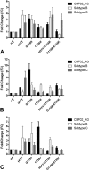
Confirmation of the role of the G118R and E138K substitutions in conferring resistance to MK-2048 in subtype C integrase. (A) Susceptibility of MK-2048-selected subtype C variant viruses to MK-2048, RAL, and EVG. (B) Dose-dependent inhibition of the strand transfer reaction of subtype C WT and G118R-, E138K-, and G118R/E138K-containing integrase proteins (INC) by the INSTI MK-2048 (0.173 nM to 5,000 nM). (C) Calculation of the FCs in IC50 values relative to WT values showing that the G118R/E138K double-variant protein has high-level in vitro resistance to MK-2048. The data presented here are the results from at least three independent experiments performed in triplicate.
FIG 4
FIG 4
Subtype-specific susceptibility of WT and variant integrase proteins to clinically relevant INSTIs. (A) DTG; (B) RAL; (C) EVG. Ki values were derived by performing strand transfer assays with variable drug (0.2 nM to 1,000 nM) and variable target DNA (0 nM to 128 nM) concentrations in the presence of fixed concentrations of LTR (160 nM) and integrase protein (300 nM). Data were fit by nonlinear regression analysis using GraphPad Prism and by a competitive inhibition equation, as detailed in Materials and Methods. FC values were calculated for each experiment by dividing the observed Ki value of each variant for a particular INSTI by that observed for the WT with the same INSTI. For each subtype, FC calculations from at least three individual experiments, analyzed by using column statistics, are presented.
FIG 5
FIG 5
Multiple-sequence alignment of subtype B, subtype C, and CRF02_AG integrase sequences from plasmids pNL4_3, pINdieC, and p97, respectively, performed by using ClustalW (v1.8). The catalytic-site residues D64, D116, and E152 are highlighted in yellow. Perfectly conserved residues are marked with asterisks, conservative substitutions are marked with : and highlighted in purple, and semiconservative substitutions are marked with • and highlighted in light blue. Nonconservative substitutions are highlighted in red. Positions E92, G140, Y143, Q148, and N155, implicated in primary resistance to RAL and EVG, are boxed in purple. Labeled residues have been adequately characterized as affecting DTG susceptibility in this study (white text with black highlighting) or in previous studies (blue boldface type) (16, 17, 37, 42).
FIG 6
FIG 6
Modeled HIV-1 WT and G118R target capture complexes showing the differential impact of G118R on the active sites of the three integrase subtypes. HIV-1 WT or G118R monomeric homology models for each of the three subtypes were created based on the structure of the freeze-trapped PFV target capture complex (PDB accession number 4E7K). These models were then used to build dimers, as described in Materials and Methods. Integrase-DNA interaction as well as cation binding in the TCC were mimicked by direct overlay of the LTR DNA, target DNA, and the Mn2+ and Zn2+ ligands from the PFV structure. The impact of the G118R substitution on the strand transfer reaction with the active site of subtype B (A), CRF02_AG (B), and subtype C (C) was analyzed by visual assessment of the differing side-chain and backbone interactions as well as residue-DNA clashes that occur with the G118R substitution, particularly within the putative strand transfer zone (yellow circles). Structural visualization and manipulation were performed by using PyMOL. Protein and DNA structures are shown as cartoons, with the residues under investigation being shown as sticks. The catalytic triad residues are labeled in blue and shown as line traces. Structures of active-site residues interacting with residues 118, 51, and 138 are shown as line traces. Possible hydrophilic interactions between atoms separated by <3.5 Å are shown by black dashed lines. Colors of lines and sticks are based on main-chain color as well as standard atomic coloration. For clarity and where necessary, the colors of residue labels are the same as those in the cartoons for that particular model.
FIG 7
FIG 7
Active-site modeling of DTG-bound integrase. For each subtype, HIV-1 WT, G118R, H51Y/G118R, or G118R/E138K monomeric models were created based on the structure of the DTG-bound PFV structure (PDB accession number 3S3M). These models were then used to build dimers as described in Materials and Methods. Integrase-DNA interaction, cation binding, and DTG-integrase interactions were mimicked by direct overlay of the LTR DNA, the Mn2+ and Zn2+ ligands, and DTG from the PFV structure reported under PDB accession number 3S3M. (A) Overlay of WT models for subtype B (green), subtype C (pink), and CRF02_AG (turquoise) showing key backbone interactions of G118. (B) Closeup view of the subtype B WT model showing interresidue and DNA-integrase interactions of H51. (C) Closeup view of the CRF02_AG WT model showing interresidue and DNA-integrase interactions of H51. (D) Closeup view of the subtype C WT model showing interresidue and DNA-integrase interactions of H51. (E) Closeup view of the subtype B WT model showing interresidue and DNA-integrase interactions of E138. (F) Closeup view of the CRF02_AG WT model showing interresidue and DNA-integrase interactions of E138. (G) Closeup view of the subtype C WT model showing interresidue and DNA-integrase interactions of E138. (H) Overlay of WT (turquoise) and G118R (yellow) active sites showing the impact of the G118R substitution on the CRF02_AG active site. Changes in the D64 side-chain orientation caused by 118R are indicated by an open blue arrow, and other key side-chain orientation changes are indicated by a solid blue arrow; the possible repositioning of at least one Mg2+ cation is indicated by a dotted red arrow. (I) Overlay of WT (pink) and G118R (purple) active sites showing the impact of the G118R substitution on the subtype C active site. Changes in the D64 side-chain orientation caused by 118R are indicated by an open blue arrow, and other key side-chain orientation changes are indicated by a solid blue arrow; the possible repositioning of at least one Mg2+ cation is indicated a the dotted red arrow. (J) Overlay of subtype B models of WT (green), G118R (tan), H51Y/G118R (red), and G118R/E138K (orange) integrase proteins showing the positioning of the three residues H51Y, G118R, and E138K relative to DTG. (K) Overlay of CRF02_AG models of the WT (turquoise), G118R (yellow), H51Y/G118R (olive green), and G118R/E138K (tan) integrase proteins showing the positioning of the three residues H51Y, G118R, and E138K relative to DTG. (L) Overlay of subtype C models of WT (pink), G118R (purple), H51Y/G118R (navy), and G118R/E138K (light blue) integrase proteins showing the positioning of the three residues H51Y, G118R, and E138K relative to DTG. Structural visualization and manipulation were performed by using PyMOL. Protein and DNA structures are shown as cartoons, with the residues under investigation being shown as sticks. The catalytic triad residues are labeled in blue and shown as line traces. Structures of active-site residues interacting with residues 118, 51, and 138 are shown as line traces. Possible hydrophilic interactions between atoms separated by <3.5 Å are shown as black dashed lines. Colors of lines and sticks are based on main-chain color as well as standard atomic coloration. For clarity and where necessary, the colors of residue labels are the same color as those in the cartoon for that particular model.

References

    1. Kommer C. 2013. New UNAIDS figures. Kinderkrankenschwester 32:78 (In German.). - PubMed
    1. International Association of Physicians in AIDS Care. 2003. HAART slashed AIDS death rates by 80 percent. IAPAC Mon 9:279. - PubMed
    1. Stephenson J. 1999. The art of ‘HAART’: researchers probe the limits of aggressive HIV treatments. JAMA 277: 614–616. doi:10.1001/jama.1997.03540320016008. - DOI - PubMed
    1. Williams A, Friedland G. 1997. Adherence, compliance, and HAART. AIDS Clin Care 9:51–54, 58. - PubMed
    1. Asahchop EL, Wainberg MA, Sloan RD, Tremblay CL. 2012. Antiviral drug resistance and the need for development of new HIV-1 reverse transcriptase inhibitors. Antimicrob Agents Chemother 56:5000–5008. doi:10.1128/AAC.00591-12. - DOI - PMC - PubMed

Publication types