

Case Reports
doi: 10.1080/08998280.2015.11929183.
Intracranial germinoma
Affiliations
- PMID: 25552796
- PMCID: PMC4264708
- DOI: 10.1080/08998280.2015.11929183
Item in Clipboard
Case Reports
Intracranial germinoma
Proc (Bayl Univ Med Cent).
2015 Jan.
Abstract
Pineal region tumors make up less than 1% of all intracranial neoplasms, with the majority being of germ cell origin. We describe the diagnostic evaluation and treatment of a patient presenting with neurological deficits who was found to have a germinoma of the pineal gland.
Figures
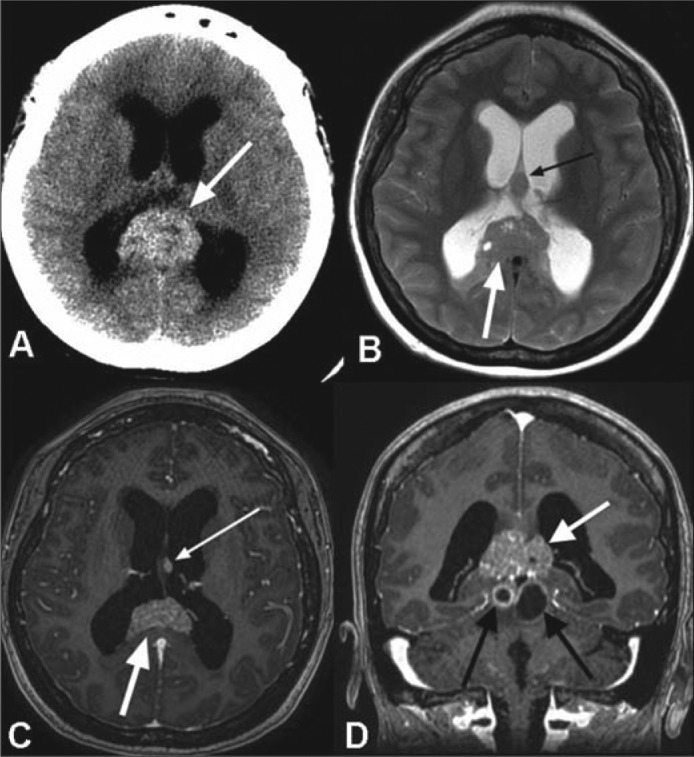
(a) Axial noncontrast CT image demonstrates a pineal region mass (white arrow) with intrinsic hyperdensity and associated obstructive hydrocephalus. (b) Axial T2-weighted, (c) axial, and (d) coronal postcontrast T1-weighted MR images reveal a T2 signal isointense to gray matter with intermixed cystic foci (white arrow in b) and avid enhancement following contrast administration (large white arrows in c and d). A separate T2 hypointense (small black arrow in b) and enhancing (small white arrow in c) nodule is seen at the interface of the septum pellucidum and left forniceal body, which suggests local cerebrospinal fluid dissemination of tumor. Additional, peripherally enhancing cystic foci are present within the midbrain tectum (black arrows in d).
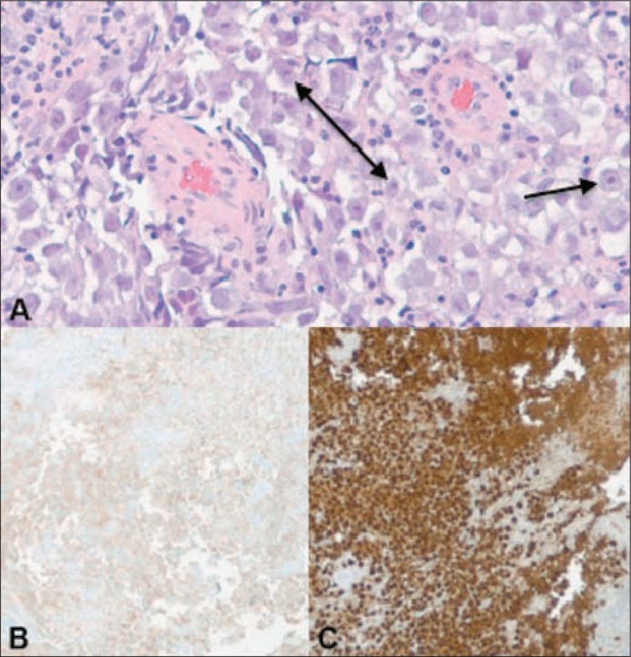
(a) Histologic examination of the biopsy specimen reveals predominantly monotonous polygonal shaped cells with prominent nucleoli and cytoplasm ranging from clear to eosinophilic. Immunohistochemical stains for (b) OCT 3/4 and (c) CD117 reveal strong reactivity. CD117 marker may also be found in other tumors such as gastrointestinal stromal tumors, whereas OCT 4 is relatively specific for germ cells. When CD117 and OCT 4 are used together, they are quite specific for a germinoma.
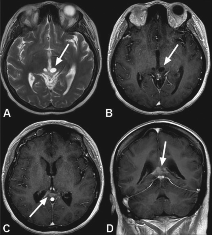
MR images following radiotherapy show a marked improvement in the appearance of the tumor bed. Near complete resolution of the large mass in the pineal fossa is seen with minimal residual enhancement along the margin of the splenium of the corpus callosum (arrows in c and d). There is considerable reduction in the extent of the previously seen peripheral enhancement involving the cystic components within the tectum of the midbrain (arrows in a and b).
References
-
- Shibui S, Nomura K. Statistical analysis of pineal tumors based on the data of Brain Tumor Registry of Japan. Prog Neurol Surg. 2009;23:1–11. - PubMed
-
- Ferla S, Spartà S, Giordano R, Zorat PL, Marin G, Meneghetti G. Pineal germinoma: diagnosis, treatment and tumor response. Ital J Neurol Sci. 1987;8(3):267–270. - PubMed
-
- Jennings MT, Gelman R, Hochberg F. Intracranial germ-cell tumors: natural history and pathogenesis. J Neurosurg. 1985;63(2):155–167. - PubMed
Publication types
LinkOut - more resources
Full Text Sources
Other Literature Sources