. 2015 Jan 1;7(1):107-16.
doi: 10.2741/E721.

ECG in neonate mice with spinal muscular atrophy allows assessment of drug efficacy

Affiliations

ECG in neonate mice with spinal muscular atrophy allows assessment of drug efficacy

Christopher R Heier et al. Front Biosci (Elite Ed). .

Abstract

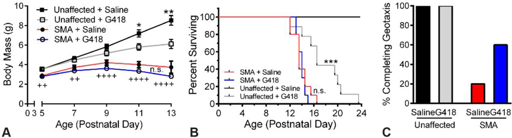
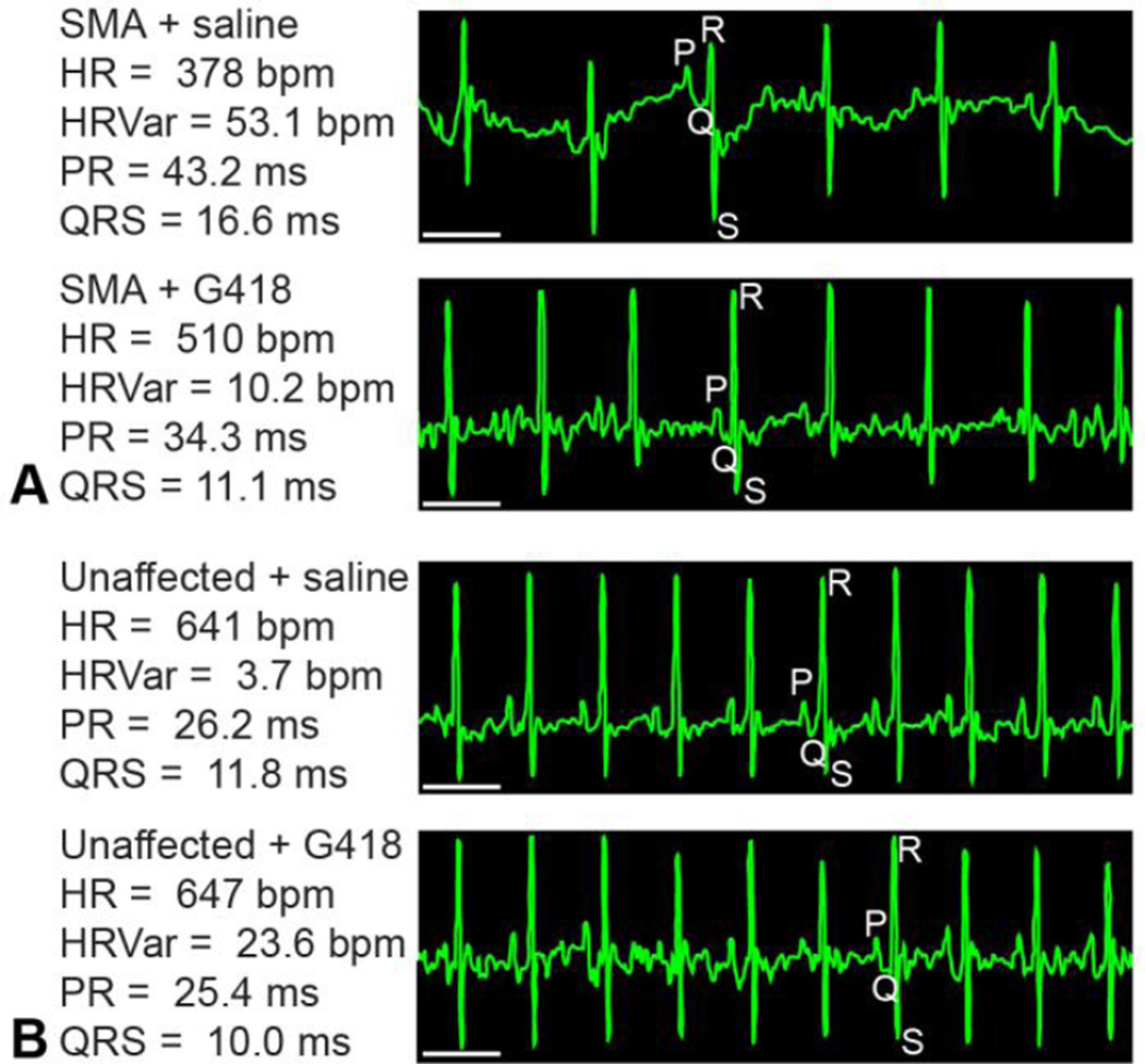
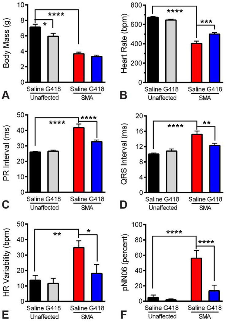
Molecular technologies have produced diverse arrays of animal models for studying genetic diseases and potential therapeutics. Many have neonatal phenotypes. Spinal muscular atrophy (SMA) is a neuromuscular disorder primarily affecting children, and is of great interest in translational medicine. The most widely used SMA mouse models require all phenotyping to be performed in neonates since they do not survive much past weaning. Pre-clinical studies in neonate mice can be hindered by toxicity and a lack of quality phenotyping assays, since many assays are invalid in pups or require subjective scoring with poor inter-rater variability. We find, however, that passive electrocardiography (ECG) recording in conscious 11-day old SMA mice provides sensitive outcome measures, detecting large differences in heart rate, cardiac conduction, and autonomic control resulting from disease. We find significant drug benefits upon treatment with G418, an aminoglycoside targeting the underlying protein deficiency, even in the absence of overt effects on growth and survival. These findings provide several quantitative physiological biomarkers for SMA preclinical studies, and will be of utility to diverse disease models featuring neonatal cardiac arrhythmias.


Figures

Figure 1
Figure 1
G418 improves motor function but not survival of SMA mice. A) Body mass was assayed from PND5 to PND13 for SMA mice and unaffected littermates receiving G418 or vehicle (n of 10, n.s. is not significant, *P less than 0.0.5, **P less than 0.0.05 for G418 versus saline, ++P less than 0.0.1, ++++P less than 0.0.001 for unaffected saline versus SMA saline). B) Kaplan-Meier survival curve of mice receiving G418 or saline vehicle (n.s. is not significant, *** P less than 0.0.005). C) Percentage of mice able to complete the negative geotaxis motor function test at PND10.
Figure 2
Figure 2
ECG of SMA and G418 treatment groups. Representative ECG waveform tracings obtained from conscious (A) SMA and (B) unaffected littermates at PND11 are presented, along with the mean waveform values for that mouse recording. Individual waveforms are denoted by P, Q, R or S. Scale bar represents 100 ms.
Figure 3
Figure 3
Quantitative ECG benefits from G418 treatment of SMA mice. A) Body mass of treatment groups at time of ECG analysis on PND11. B) Heart rate of treatment groups, showing SMA bradycardia and effects of drug treatment. C) PR interval, showing heart block in SMA mice and effects of drug treatment. D) QRS interval, showing cardiac conduction deficits in SMA mice and effects of drug treatment. E–F) Measures of heart rate variability, showing alteration of HR Variability (E) and pNN06 (F) in SMA mice and the effects of drug treatment. (n of 10, * P less than 0.0.5, ** P less than 0.0.1, *** P less than 0.0.005, **** P less than 0.0.001)
Figure 4
Figure 4
ECG at end stages of G418 toxicity. ECGs were recorded in mice of unaffected genotypes at advanced stages of G418 toxicity, along with littermate saline controls. A) Body mass of G418 and saline injected mice. B) Heart rates and (C) PR intervals were affected by G418 toxicity. D) QRS interval was not significantly affected by toxicity. E–F) Measures of heart rate variability, reflecting autonomic nerve control of the heart, were not affected by toxicity. (n of 4, ages range from PND19 to PND23, * P less than 0.0.5)

References

    1. Lefebvre S, Burglen L, Reboullet S, Clermont O, Burlet P, Viollet L, Benichou B, Cruaud C, Millasseau P, Zeviani M, Le Paslier D, Frezal J, Cohen D, Weissenbach J, Munnich A, Melki J. Identification and characterization of a spinal muscular atrophy-determining gene. Cell. 1995;80(1):155–165. - PubMed
    1. Wirth B. An update of the mutation spectrum of the survival motor neuron gene (SMN1) in autosomal recessive spinal muscular atrophy (SMA) Hum Mutat. 2000;15(3):228–237. - PubMed
    1. Rochette CF, Gilbert N, Simard LR. SMN gene duplication and the emergence of the SMN2 gene occurred in distinct hominids: SMN2 is unique to Homo sapiens. Hum Genet. 2001;108(3):255–266. - PubMed
    1. Lorson CL, Hahnen E, Androphy EJ, Wirth B. A single nucleotide in the SMN gene regulates splicing and is responsible for spinal muscular atrophy. Proc Natl Acad Sci U S A. 1999;96(11):6307–6311. - PMC - PubMed
    1. Feldkotter M, Schwarzer V, Wirth R, Wienker TF, Wirth B. Quantitative analyses of SMN1 and SMN2 based on real-time lightCycler PCR: fast and highly reliable carrier testing and prediction of severity of spinal muscular atrophy. Am J Hum Genet. 2002;70(2):358–368. - PMC - PubMed

Publication types

MeSH terms