

Spontaneous cervical-mediastinal hematoma caused by hemorrhage into parathyroid adenoma: A clinical case
- PMID: 25553526
- PMCID: PMC4334879
- DOI: 10.1016/j.ijscr.2014.10.029
Spontaneous cervical-mediastinal hematoma caused by hemorrhage into parathyroid adenoma: A clinical case
Abstract
Introduction: Spontaneous cervical-mediastinal hematoma caused by extracapsular rupture of parathyroid gland occurs extremely rarely. There are no standard treatment approaches because of the peculiarities of each case.
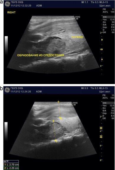
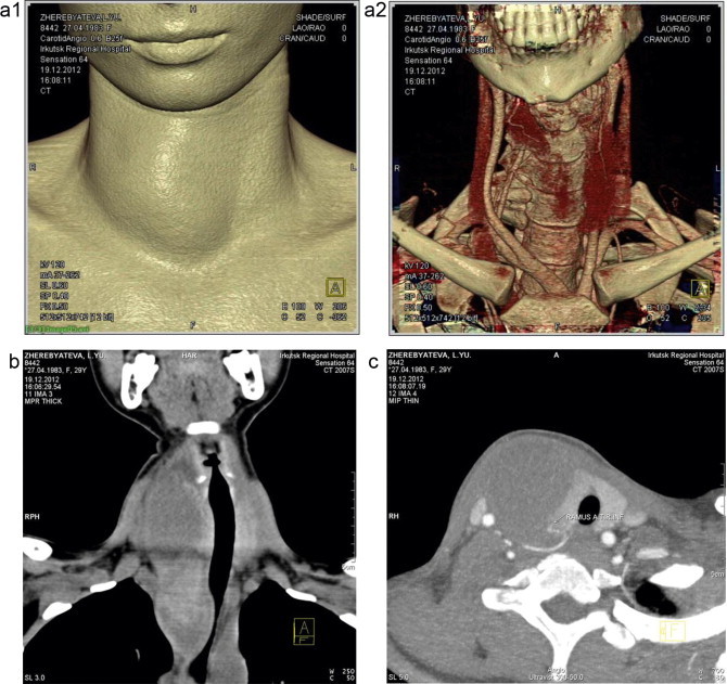
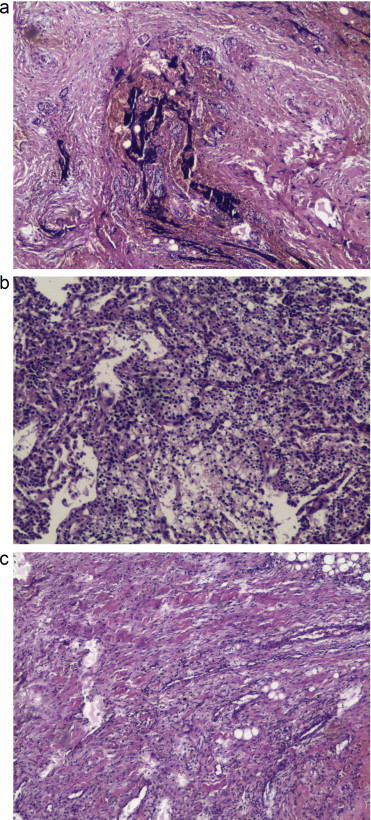
Presentation of case: We report herewith about a rare case of spontaneous cervical-mediastenal hematoma occured by hemorrhage in parathyroid adenoma, which was detected in an previously absolutely healthy female patient in the age of 29. This woman was hospitalized in 2 days after the manifestation, complaining about a neck ache. Indirect laryngoscopy: right-side larynx paresis. Blood test: parathyroid hormone 843pg/ml (norm 15-65), ionized calcium 1.8mmol/l (norm 0.9-1.1). Positive dynamics was observed throughout 8 days of anti-inflammatory therapy. Symptoms of neck organs compression increased acutely at the 9th day. The patient was operated - hematoma lancing with resection of walls. Histological examination discovered the fragments of parathyroid adenoma in the hematoma's wall. Level of ionized blood calcium got normal approximately in 24h after the surgery. The patient was examined 6 months after the surgery. The patient had no disphagy, voice quality was intact, breathing was not restricted. Level of parathyroid hormone in blood got normal.
Discussion: A rareness of this pathology and treatment variability does not allow to choose a unified medical and diagnostic tactics.
Conclusion: Our case demonstrates that radical correction of primary hyperparathyroidism by excision of hematoma and its fibrous capsule with preservation of thyroid gland is possible in conditions of tense cervical-mediastinal hematoma with inflammation process in the hemorrhage area.
Keywords: Acute neck diseases; Extracapsular parathyroid hemorrhage; Hypercalcemia; Hyperparathyroid gland disease; Larynx paresis; Tumors of parathyroid glands.
Copyright © 2014 The Authors. Published by Elsevier Ltd.. All rights reserved.
Figures
References
-
- Nito T., Miyajima C., Kimura M., Sugasawa M. Parathyroid adenoma causing spontaneous cervical hematoma: a case report. Acta Otolaryngol Suppl. 2007:160–163. Dec;(559) - PubMed
-
- Chaffanjon P.C., Chavanis N., Chabre O., Brichon P.Y. Extracapsular hematoma of the parathyroid glands. World J Surg. 2003;27(1):14–17. [abstract] - PubMed
-
- Il’icheva E.A., Shpakova E.A., Roi T.A., Makhutov V.N., Tarnueva I.F., Moshkova E.S. Specific features of laryngeal paresis following surgical treatment of diffuse toxic goiter (a prospective longitudinal passive study) Vestn Otorinolaringol. 2011;3:51–54. [abstract] - PubMed
LinkOut - more resources
Full Text Sources
Other Literature Sources
Research Materials
