Comparative Study
. 2014 Nov;6(11):957-74.
doi: 10.18632/aging.100705.

Age-associated changes in basal NF-κB function in human CD4+ T lymphocytes via dysregulation of PI3 kinase

Affiliations
Comparative Study

Age-associated changes in basal NF-κB function in human CD4+ T lymphocytes via dysregulation of PI3 kinase

Arsun Bektas et al. Aging (Albany NY). 2014 Nov.

Abstract

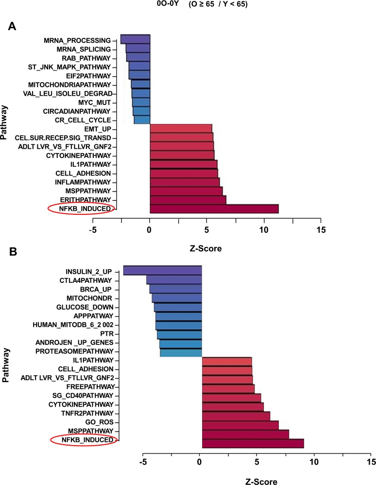
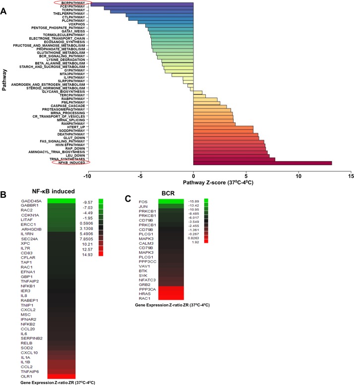
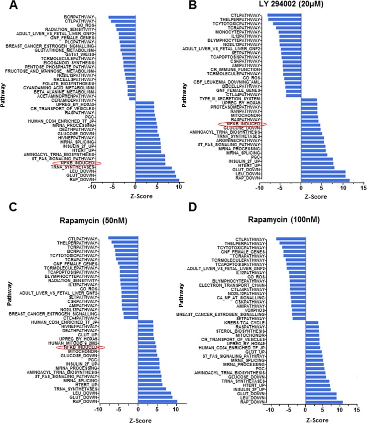
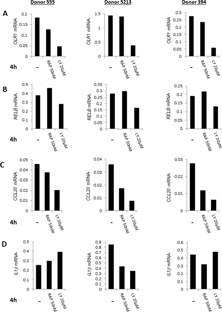
Immune impairment and high circulating level of pro-inflammatory cytokines are landmarks of human aging. However, the molecular basis of immune dys-regulation and the source of inflammatory markers remain unclear. Here we demonstrate that in the absence of overt cell stimulation gene expression mediated by the transcription factor NF-κB is higher in purified and rested human CD4+ T lymphocytes from older compared to younger individuals. This increase of NF-κB -associated transcription includes transcripts for pro-inflammatory cytokines such as IL-1 and chemokines such as CCL2 and CXCL10. We demonstrate that NF-κB up-regulation is cell-intrinsic and mediated in part by phosphatidylinositol 3-kinase (PI3K) activity induced in response to metabolic activity, which can be moderated by rapamycin treatment. Our observations provide direct evidence that dys-regulated basal NF-κB activity may contribute to the mild pro-inflammatory state of aging.


Conflict of interest statement

Conflict of interest statement

The authors of this manuscript have no conflict of interest to declare.

Figures

Figure 1
Figure 1. Effect of age on gene expression profiling in unactivated human peripheral blood CD4+ T lymphocytes
Total RNA isolated from CD4+ T cells that had been incubated for 4h at 37°C was converted to biotin labeled cRNA and hybridized to Illumina HumanRef-8 Expression BeadChip. Normalized hybridization data was analyzed using Parametric Analysis of Gene Set Enrichment (PAGE) (Broad Institute, M.I.T., Cambridge MA). (A) a set of 31 donors with age range 25-81 (Table S1A), (B) an independent set of 23 donors with age range 26-83 (Table S1B). Pathway Z-scores were averaged amongst less than 65 (Y) and 65 and older (O) individuals and the Z-score difference between O and Y is shown on the X-axis. Each row denotes a different pathway (p-value ≤ 0.05 and fdr ≤ 0.3).
Figure 2
Figure 2. Effect of increased metabolic activity on gene expression profile in CD4+ T lymphocytes
RNA samples from 23 donors (age range 29-82) (Table S1C) were obtained from freshly purified CD4+ T cells (maintained at 4°C) or from cells incubated at 37°C for 4h. The 46 samples were converted to biotin labeled cRNA and hybridized to Illumina HumanRef-8 Expression BeadChip. (A) Normalized data were analyzed by PAGE and the average Z-score for individual pathways compared between 4°C samples and those incubated at 37°C to obtain a Z-score difference x-axis; Z-score (37°C-4°C ). Rows denote individual pathways. Most prominently up- and down-regulated pathways at 37°C are indicated in red. (B) and (C) Statistically significant genes within the NF-κB induce (up-regulated) and BCR (down-regulated) pathways.
Figure 3
Figure 3. Gene expression following pharmacologic perturbation of PI3K in CD4+ T lymphocytes
CD4+ T cells were incubated at 37°C for 4h in the absence of pharmacologic agents or in the presence of LY 294002 (20μM) or rapamycin (50nM or 100nM, as indicated). RNA isolated from freshly purified cells or cells treated as above was converted to biotin labelled cRNA and hybridized to Illumina HumanRef-8 Expression BeadChip. Normalized data were analyzed by PAGE and the average Z-score for individual pathways compared between fresh samples (maintained at 4°C and (A) cells incubated in the absence of inhibitors, (B) cells incubated in the presence of 20μM LY 294002, (C) cells incubated in the presence of 50nM rapamycin and (D) cells incubated in the presence of 100 nM rapamycin. Rows denote individual pathways.
Figure 4
Figure 4. Quantative RT-PCR validation of the effects of pharmacologic agents on gene expression in CD4+ T lymphocytes
CD4+ T cells from three donors (ages 30, 63, 73) were cultured at 37°C for 4h in the presence of LY294002 (20μM), rapamycin (50nM) or in the absence of added drugs. RNA prepared from these cells was assayed for expression of (A) OLR1, (B) RELB, (C) CCL20 and (D) IL1β by quantitative RT-PCR. Expression values after normalization to GAPDH are shown on the Y axis.

References

    1. Bruunsgaard H, Pedersen BK. Age-related inflammatory cytokines and disease. Immunol Allergy Clin North Am. 2003;23:15–39. - PubMed
    1. Ferrucci L, Corsi A, Lauretani F, Bandinelli S, Bartali B, Taub DD, Guralnik JM, Longo DL. The origins of age-related proinflammatory state. Blood. 2005;105:2294–2299. - PMC - PubMed
    1. Pawelec G. Hallmarks of human "immunosenescence": adaptation or dysregulation? Immunity & ageing : I & A. 2012;9:15. - PMC - PubMed
    1. Goronzy JJ, Weyand CM. Understanding immunosenescence to improve responses to vaccines. Nature immunology. 2013;14:428–436. - PMC - PubMed
    1. Pawelec G, Akbar A, Caruso C, Solana R, Grubeck-Loebenstein B, Wikby A. Human immunosenescence: is it infectious? Immunol Rev. 2005;205:257–268. - PubMed

Publication types

MeSH terms