. 2015 Jan 2;347(6217):67-71.
doi: 10.1126/science.1260972.

Innate immunity. Dermal adipocytes protect against invasive Staphylococcus aureus skin infection

Affiliations

Innate immunity. Dermal adipocytes protect against invasive Staphylococcus aureus skin infection

Ling-juan Zhang et al. Science. .

Abstract

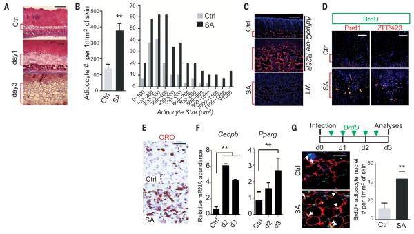
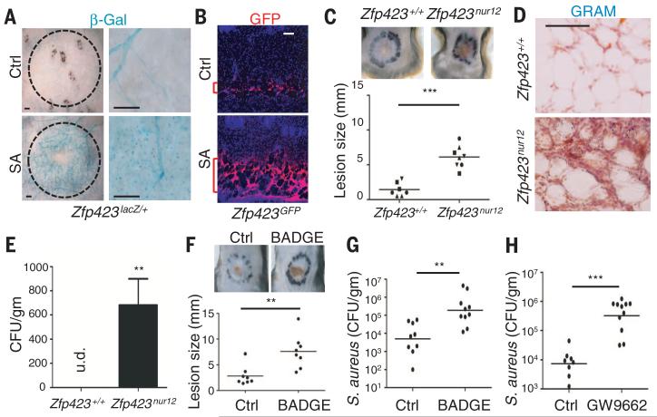
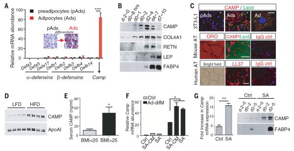
Adipocytes have been suggested to be immunologically active, but their role in host defense is unclear. We observed rapid proliferation of preadipocytes and expansion of the dermal fat layer after infection of the skin by Staphylococcus aureus. Impaired adipogenesis resulted in increased infection as seen in Zfp423(nur12) mice or in mice given inhibitors of peroxisome proliferator-activated receptor γ. This host defense function was mediated through the production of cathelicidin antimicrobial peptide from adipocytes because cathelicidin expression was decreased by inhibition of adipogenesis, and adipocytes from Camp(-/-) mice lost the capacity to inhibit bacterial growth. Together, these findings show that the production of an antimicrobial peptide by adipocytes is an important element for protection against S. aureus infection of the skin.


Figures

Fig. 1
Fig. 1. Skin infection stimulates an increase in dermal adipocytes
(A) Hematoxylin and eosin staining of mouse skin injected with phosphate-buffered saline (PBS) control (Ctrl) or S. aureus (SA). Red brackets indicate subcutaneous adipose layer. Scale bars, 200 μm. (B) Quantification of the number and size distribution of Caveolin+/Perilipin+ adipocytes 3 days after Ctrl or S. aureus injection (n = 3 to ~5 mice/group and 3 microscopy fields/mice). (C) Increase of adiponectin positive cells seen by staining for β-Gal (red) 3 days after S. aureus or Ctrl injection in AdipoQcre;R26R mice. Wild-type mice injected with S. aureus are shown as a staining control. Scale bars, 200 μm. Nuclei were stained with 4′,6-diamidino-2-phenylindole (DAPI) (blue). (D) Ctrl or S. aureus–infected mice (day 1) were injected with BrdU 4 hours before analysis. Proliferative preadipocytes were identified by means of yellow staining showing colocalization of PREF1 or ZFP423 (red) and BrdU (green). Scale bars, 100 μm. (E) Dermal cells were isolated from Ctrl or S. aureus (SA)–infected skin then treated with adipocyte differentiation medium for 5 days. Lipid production was shown by Oil-Red-O (ORO) staining. Scale bars, 200 μm. (F) Relative mRNA expression for Cebpb and Pparg (n = 3 mice/group) in skin after S. aureus infection. (G) (Top) Schematic of 3-day BrdU labeling experiments during S. aureus infection. (Left) Representative images for caveolin (red) and BrdU (green) staining of skin sections. Arrows indicate Caveolin+BrdU+ adipocytes. Scale bars, 50 μm. (Right) Quantification of the number of Caveolin+BrdU+ adipocytes (n = 3~5 mice/group and 3 microscopy fields/mice). All error bars indicate mean ± SEM; ** P < 0.01 (t test).
Fig. 2
Fig. 2. Adipocytes are essential for host defense against S. aureus infection
(A) β-gal staining of the underside of Zfp423lacZ/+ skin 3 days after injection with PBS (Ctrl) or S. aureus. Injected area is indicated by dotted line. Scale bars, 1mm (left), 200 μm (right). (B) GFP immunostaining (red) of skin sections from Ctrl or S. aureus–injected Zfp423GFP mice. Scale bars, 100 μm. (C) Infected lesion size in Zfp423nur12 or Zfp423+/+ mice (n = 7~8/group) and (D) bacteria in adipose observed by Gram staining (dark blue). Scale bars, 100 μm. (E) Systemic bacteremia detected 3 days after S. aureus skin injection in spleens isolated from Zfp423nur12 mice but not Zfp423+/+ mice (n = 3 mice/group). Scale bars, 200 μm. (F to H) BADGE or GW9662 increased susceptibility to S. aureus as observed by (F) increased lesion size and [(G) and (H)] increased S. aureus (CFU) from the skin calculated with plate counting (n = 8 to ~10/group). All error bars indicate mean ± SEM. ** P < 0.01, *** P < 0.001 (t test).
Fig. 3
Fig. 3. Adipocytes produce cathelicidin antimicrobial peptide
(A) 3T3-L1 preadipocytes (pAds) were differentiated to adipocytes (Ads). Expression of mRNA for α-defensins, β-defensins, and cathelicidin (Camp) is shown (n = 3 cultures/group). (Inset) Oil-Red-O staining. (B) Western-blotting of conditioned media from differentiating 3T3-L1 cells. (C) Staining of 3T3-L1 cells or mouse and human subcutaneous adipose tissue (AT) by using lipid dye (Bodipy; green), mouse CAMP (red), Oil-red-O, or the human cathelicidin peptide LL37 (red) as indicated. Nuclei are counterstained with DAPI. Scale bar, 5 μm (3T3-L1) or 50 μm (adipose tissue). (D) C57BL6 mice fed with low-fat diet (LFD) or high-fat diet (HFD) for 4 weeks, and mouse serum was collected for Western blotting analysis by using CAMP and ApoAI antibodies. (E) Human serum cathelicidin measured in overweight (/BMI >25) or normal-weight human subjects (n = 10 subjects/group). (F) Camp mRNA expression levels were examined by quantitative reverse transcription PCR (RT-PCR) in 3T3L1 preadipocytes treated with or without adipocyte differentiation (Ad-difM) medium and with S. aureus–conditioned medium (SA-CM) or UV-killed S. aureus (SA). (G) Primary cultures of dermal cells isolated from Ctrl or S. aureus–injected skin were treated with Ad-difM. Relative Camp mRNA fold induction at day 2 compared with undifferentiated control were examined by means of quatitative (RT-PCR) (left); CAMP and FABP4 protein were detected in conditioned medium by means of Western blot (right). All error bars indicate mean ± SEM; *P < 0.05, **P < 0.01, ***P < 0.001 (t test).
Fig. 4
Fig. 4. Adipocytes kill bacteria by producing cathelicidin
(A) CAMP immunostaining (left) of adipose in mice infected with S. aureus. Scale bar, 100 μm. (B) Western-blotting of CAMP from whole-cell extracts (WCE) or conditioned medium (CM) from 3T3-L1 pAds or Ads. Red arrow indicates mature peptide form, and red asterisks indicate precursor forms of cathelicidin. Synthetic CRAMP peptide is shown as standard for the processed form of CAMP in neutrophils. (C) Growth curve of S. aureus in Ctrl medium or conditioned medium (CM) from pAd or Ad as indicated. (Inset) CAMP immunoblot in conditioned medium. (D) S. aureus growth in media alone (Ctrl) or with addition of protein extracted from subcutaneous adipose tissue from Camp+/+ or Camp−/− mice (n = 3). (E) CAMP expression is suppressed in adipose in Zfp423-deficient mice. Scale bars, 100 μm. (F) Camp mRNA expression in S. aureus– or PBS-injected Zfp423+/+ and Zfp423nur12 mice (n = 5 mice/group). (G) Camp+/+ or Camp−/− mice were pretreated with BADGE or dimethyl sulfoxide (ctrl) before infecting with S. aureus and S. aureus survival in skin measured 3 days later (n = 7 to ~10 mice/group). All error bars indicate mean ± SEM. *P < 0.05, **P < 0.01, ***P < 0.001 (one-way analysis of variance). Red brackets indicate subcutaneous adipose layer.

Comment in

Similar articles

Cited by

References

    1. Nestle FO, Di Meglio P, Qin JZ, Nickoloff BJ. Nat. Rev. Immunol. 2009;9:679–691. - PMC - PubMed
    1. Gallo RL, Hooper LV. Nat. Rev. Immunol. 2012;12:503–516. - PMC - PubMed
    1. Nizet V, et al. Nature. 2001;414:454–457. - PubMed
    1. Ong PY, et al. N. Engl. J. Med. 2002;347:1151–1160. - PubMed
    1. Pütsep K, Carlsson G, Boman HG, Andersson M. Lancet. 2002;360:1144–1149. - PubMed

Publication types

MeSH terms