. 2015 Feb 1:126:169-77.
doi: 10.1016/j.colsurfb.2014.12.020. Epub 2014 Dec 16.

Serum protein layers on parylene-C and silicon oxide: effect on cell adhesion

Affiliations

Serum protein layers on parylene-C and silicon oxide: effect on cell adhesion

Evangelos Delivopoulos et al. Colloids Surf B Biointerfaces. .

Abstract

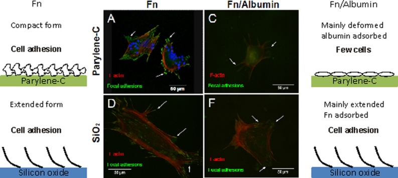
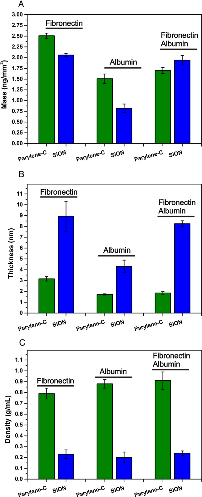
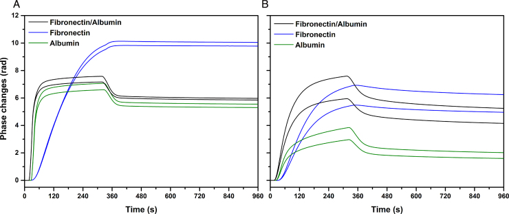
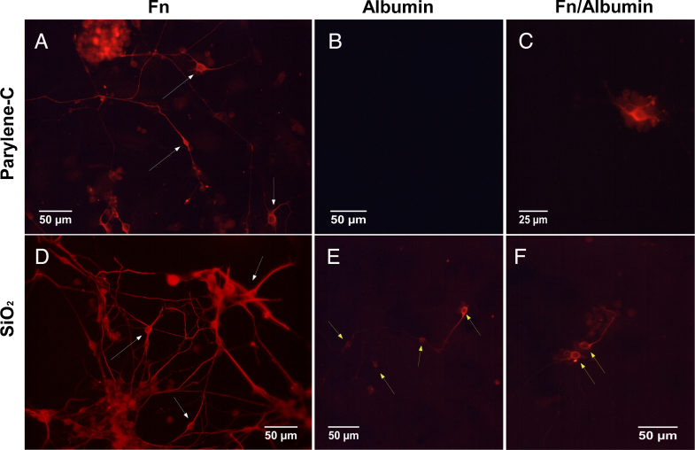
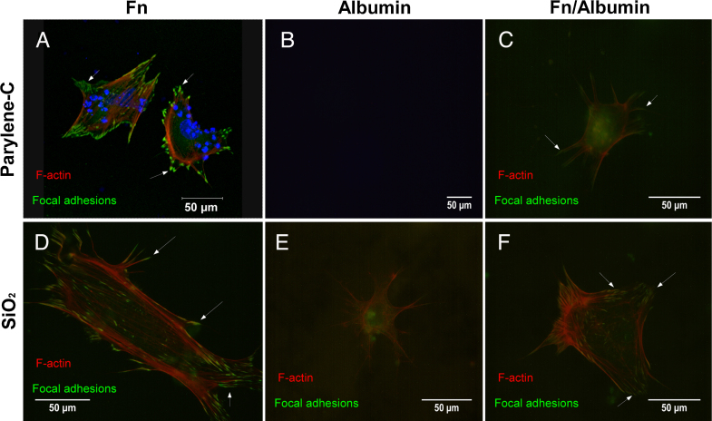
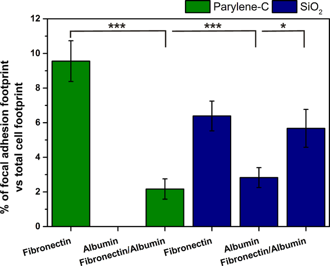
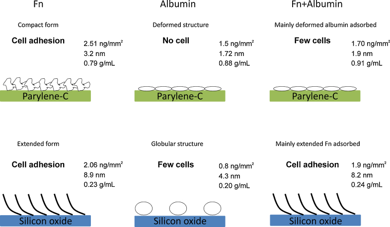
Among the range of materials used in bioengineering, parylene-C has been used in combination with silicon oxide and in presence of the serum proteins, in cell patterning. However, the structural properties of adsorbed serum proteins on these substrates still remain elusive. In this study, we use an optical biosensing technique to decipher the properties of fibronectin (Fn) and serum albumin adsorbed on parylene-C and silicon oxide substrates. Our results show the formation of layers with distinct structural and adhesive properties. Thin, dense layers are formed on parylene-C, whereas thicker, more diffuse layers are formed on silicon oxide. These results suggest that Fn acquires a compact structure on parylene-C and a more extended structure on silicon oxide. Nonetheless, parylene-C and silicon oxide substrates coated with Fn host cell populations that exhibit focal adhesion complexes and good cell attachment. Albumin adopts a deformed structure on parylene-C and a globular structure on silicon oxide, and does not support significant cell attachment on either surface. Interestingly, the co-incubation of Fn and albumin at the ratio found in serum, results in the preferential adsorption of albumin on parylene-C and Fn on silicon oxide. This finding is supported by the exclusive formation of focal adhesion complexes in differentiated mouse embryonic stem cells (CGR8), cultured on Fn/albumin coated silicon oxide, but not on parylene-C. The detailed information provided in this study on the distinct properties of layers of serum proteins on substrates such as parylene-C and silicon oxide is highly significant in developing methods for cell patterning.

Keywords: Biosensing technique; Cell adhesion; Fibronectin; Parylene-C; Serum protein adsorption; Silicon oxide.


Figures

None
Graphical abstract
Fig. 1
Fig. 1
Real time TM and TE phase changes for adsorption of Fn at 50 μg/mL (A) and albumin at 5000 μg/mL (B) on parylene-C and SiON in PBS at 20 °C.
Fig. 2
Fig. 2
Surface coverage (ng/mm2) (A), thickness (nm) (B) and density (g/mL) (C) of adsorbed Fn (50 μg/mL), albumin (5000 μg/mL) and mixture of Fn/albumin (50 μg/mL/5000 μg/L) on parylene-C and SiON in PBS at 20 °C. Data is expressed as mean ± SD (n = 3).
Fig. 3
Fig. 3
Real time TM and TE phase changes for adsorption on parylene-C (A) and SiON (B) of a mixture of Fn at 50 μg/mL and albumin at 5000 μg/mL compared with Fn at 50 μg/mL and albumin at 5000 μg/mL in PBS at 20 °C (injection start times are offset for clarity).
Fig. 4
Fig. 4
Representative field of views of neuronal populations from differentiated CGR8 cultures on protein treated parylene-C and SiO2 substrates. The red fluorescence denotes b-tubulin III on Fn (A), albumin (B) and Fn/albumin (C) coated parylene-C; Fn (D), albumin (E) and Fn/albumin (F) coated SiO2. Fn coated parylene-C and SiO2 hosted many neurons extending long processes (white arrows in (A) and (D)), whereas no cells were present on the albumin coated parylene-C (B). Neurons on Fn/albumin coated parylene-C were sparse and did not extend processes (C). Sparse populations of neurons were also found across albumin (E) and Fn/albumin (F) coated SiO2 but they have short neuronal processes (yellow arrows). (For interpretation of the references to color in this figure legend, the reader is referred to the web version of this article.)
Fig. 5
Fig. 5
Fibroblast-like cells differentiated from mES cells (CGR8) on protein treated parylene-C and SiO2 substrates. Red, green and blue fluorescence denote F-actin, vinculin and nuclear (DAPI) staining, respectively on Fn (A), albumin (B) and Fn/albumin (C) coated parylene-C; Fn (D), albumin (E) and Fn/albumin (F) coated SiO2. Focal adhesions (white arrows) are more pronounced in Fn coated parylene-C and SiO2 (A and D) and appear as elongated areas of green fluorescence. No cells were detected on albumin coated parylene-C (B). On albumin coated SiO2 (E) vinculin is diffused throughout the cytoplasm, which denotes the absence of focal adhesions and lack of cell attachment to the ECM. The co-adsorption of Fn and albumin on parylene-C (C) and SiO2 (F) results in focal adhesion formation only on the SiO2, due to the absence of Fn from parylene-C.
Fig. 6
Fig. 6
Percentage of total focal adhesion complex area out of total cell area, for each protein coated surface. On albumin coated parylene-C no cells were detected. Two independent experiments were conducted and averages were calculated from nine cells on four different substrates, for each condition and material. Error bars denote the standard error of means (SEM). One and three dots represent statistical significance of P ≤ 0.05 and P ≤ 0.001, respectively.
Fig. 7
Fig. 7
Proposed model of the layers formed by serum proteins adsorbed onto silicon oxide and parylene-C. Fn forms a dense and thin layer on parylene-C with proteins in a compact structure that promotes cell adhesion. On the opposite, on silicon oxide, a more diffuse and thicker layer is formed suggesting that Fn adopts an extended structure that also mediates cell adhesion. Albumin forms a dense and thin layer on parylene-C with protein molecules probably deformed, whereas a globular structure is observed on silicon oxide leading to the formation of a diffuse and thicker layer. The albumin coated parylene-C prevents cell adhesion, whereas few cells can grow on the albumin coated silicon oxide. Finally, the binary mixture of Fn and albumin results in the preferential adsorption of one of the protein, deformed albumin on parylene-C and extended Fn on silicon oxide. This conclusion is supported by the significant cell adhesion process observed on silicon oxide in opposition to parylene-C.

References

    1. Herrault F., Yorish S., Crittenden T.M., Chang-Hyeon J., Allen M.G. Parylene-insulated ultradense microfabricated coils. J. Microelectromech. Syst. 2010;19:1277–1283.
    1. Temiz Y., Ferretti A., Leblebici Y., Guiducci C. A comparative study on fabrication techniques for on-chip microelectrodes. Lab Chip. 2012;12:4920–4928. - PubMed
    1. Goda T., Konno T., Takai M., Ishihara K. Photoinduced phospholipid polymer grafting on parylene film: advanced lubrication and antibiofouling properties. Colloids Surf. B: Biointerfaces. 2007;54:67–73. - PubMed
    1. Jui-Mei H., Rieth L., Normann R.A., Tathireddy P., Solzbacher F. Encapsulation of an integrated neural interface device with parylene C. IEEE Trans. Biomed. Eng. 2009;56:23–29. - PubMed
    1. Sharma A., Rieth L., Tathireddy P., Harrison R., Oppermann H., Klein M., Topper M., Jung E., Normann R., Clark G., Solzbacher F. Long term in vitro functional stability and recording longevity of fully integrated wireless neural interfaces based on the Utah Slant Electrode Array. J. Neural Eng. 2011;8:045004. - PMC - PubMed

Publication types