. 2015 Feb;125(2):782-6.
doi: 10.1172/JCI78361. Epub 2015 Jan 2.

Neuroepithelial circuit formed by innervation of sensory enteroendocrine cells

Neuroepithelial circuit formed by innervation of sensory enteroendocrine cells

Diego V Bohórquez et al. J Clin Invest. 2015 Feb.

Abstract

Satiety and other core physiological functions are modulated by sensory signals arising from the surface of the gut. Luminal nutrients and bacteria stimulate epithelial biosensors called enteroendocrine cells. Despite being electrically excitable, enteroendocrine cells are generally thought to communicate indirectly with nerves through hormone secretion and not through direct cell-nerve contact. However, we recently uncovered in intestinal enteroendocrine cells a cytoplasmic process that we named neuropod. Here, we determined that neuropods provide a direct connection between enteroendocrine cells and neurons innervating the small intestine and colon. Using cell-specific transgenic mice to study neural circuits, we found that enteroendocrine cells have the necessary elements for neurotransmission, including expression of genes that encode pre-, post-, and transsynaptic proteins. This neuroepithelial circuit was reconstituted in vitro by coculturing single enteroendocrine cells with sensory neurons. We used a monosynaptic rabies virus to define the circuit's functional connectivity in vivo and determined that delivery of this neurotropic virus into the colon lumen resulted in the infection of mucosal nerves through enteroendocrine cells. This neuroepithelial circuit can serve as both a sensory conduit for food and gut microbes to interact with the nervous system and a portal for viruses to enter the enteric and central nervous systems.


Figures

Figure 3
Figure 3. Enteroendocrine cell–neuron circuit revealed by monosynaptic rabies virus.
(A) When delivered by enema into the lumen of the colon of wild-type mice, SADΔG-GFP (ΔG-GFP) rabies infects enteroendocrine cells, as confirmed by their PYY immunoreactivity. (B) To enable the monosynaptic rabies to spread, we engineered rabG glycoprotein in PYY-containing enteroendocrine cells by breeding Pyy-Cre with RɸGT mice. (C) When delivered into the colon of Pyy-Cre–rabG (Pyy-rabG) mice, SADΔG-GFP infected enteroendocrine cells (dotted lines) and underlying connecting nerve fibers. These immunoreacted with the neuronal marker PGP 9.5. (D) Model of afferent and efferent innervation of enteroendocrine cells. Scale bars: 10 μm.
Figure 2
Figure 2. Enteroendocrine cells express pre- and postsynaptic proteins.
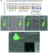
(A) Gene expression analysis of FAC-sorted Pyy-GFP enteroendocrine cells shows their expression of presynaptic and postsynaptic proteins. Fold expression represents log10 2–[(ΔCt Pyy-GFP–positive cells) (ΔCt GFP-negative epithelial cells)] and error bars represent the SEM. Significant differences were separated by Student’s t test using 2–(ΔCt) values at α = 0.05. (B) The presynaptic marker synapsin 1 (SYN1) immunoreacted with 96.4% (SEM ± 0.5%) of Pyy-GFP cells. (C) 35.3% (SEM ± 3.5%) of Pyy-GFP cells immunoreacted with the postsynaptic marker PSD95. (D) In vitro, Cck-GFP cells connecting to trigeminal neurons expressed the postsynaptic marker PSD95 (cyan). Blue, DAPI nuclear stain. Scale bars: 10 μm.
Figure 1
Figure 1. Enteroendocrine cells connect to sensory neurons in vivo and in vitro.
(AC) Confocal z-stacks were reconstructed using Imaris (Bitplane Inc.). Cck-GFP and Pyy-GFP enteroendocrine cells have a neuropod through which they contact to nerve fibers. (A) A neurofilament-medium (NfM) (red) nerve connects to an intestinal Pyy-GFP cell (green). (B) Calbindin (Calb) nerve innervates the neuropod of a colonic Pyy-GFP cell. (C) Colonic Pyy-GFP cell extends its neuropod to connect to neurofilament and CGRP nerves. (D) Coculture scheme of enteroendocrine cell and primary sensory neurons. EEC, enteroendocrine cell; TG, trigeminal neuron. (E) Time-lapse sequence showing how a single Cck-GFP enteroendocrine cell (green) connects to a sensory neuron (DiI-labeled, red) in vitro. Footage is presented in Supplemental Videos 1 and 2. (F) The enteroendocrine cell–neuron connection is stable at 23 minutes, 14 seconds, and cells remained connected for 88 hours until the end of the experiment. Time, hours:minutes. Scale bars: 10 μm.

References

    1. Critchley HD, Harrison NA. Visceral influences on brain and behavior. Neuron. 2013;77(4):624–638. doi: 10.1016/j.neuron.2013.02.008. - DOI - PubMed
    1. Furness JB, Rivera LR, Cho HJ, Bravo DM, Callaghan B. The gut as a sensory organ. Nat Rev Gastroenterol Hepatol. 2013;10(12):729–740. - PubMed
    1. Mellitzer G, et al. Loss of enteroendocrine cells in mice alters lipid absorption and glucose homeostasis and impairs postnatal survival. J Clin Invest. 2010;120(5):1708–1721. doi: 10.1172/JCI40794. - DOI - PMC - PubMed
    1. Samuel BS, et al. Effects of the gut microbiota on host adiposity are modulated by the short-chain fatty-acid binding G protein-coupled receptor, Gpr41. Proc Natl Acad Sci U S A. 2008;105(43):16767–16772. doi: 10.1073/pnas.0808567105. - DOI - PMC - PubMed
    1. Batterham RL, et al. Gut hormone PYY(3-36) physiologically inhibits food intake. Nature. 2002;418(6898):650–654. doi: 10.1038/nature00887. - DOI - PubMed

Publication types