. 2015 Jan 2;10(1):e116416.
doi: 10.1371/journal.pone.0116416. eCollection 2015.

Heterochrony and early left-right asymmetry in the development of the cardiorespiratory system of snakes

Affiliations

Heterochrony and early left-right asymmetry in the development of the cardiorespiratory system of snakes

Benjamin J van Soldt et al. PLoS One. .

Abstract

Snake lungs show a remarkable diversity of organ asymmetries. The right lung is always fully developed, while the left lung is either absent, vestigial, or well-developed (but smaller than the right). A 'tracheal lung' is present in some taxa. These asymmetries are reflected in the pulmonary arteries. Lung asymmetry is known to appear at early stages of development in Thamnophis radix and Natrix natrix. Unfortunately, there is no developmental data on snakes with a well-developed or absent left lung. We examine the adult and developmental morphology of the lung and pulmonary arteries in the snakes Python curtus breitensteini, Pantherophis guttata guttata, Elaphe obsoleta spiloides, Calloselasma rhodostoma and Causus rhombeatus using gross dissection, MicroCT scanning and 3D reconstruction. We find that the right and tracheal lung develop similarly in these species. By contrast, the left lung either: (1) fails to develop; (2) elongates more slowly and aborts early without (2a) or with (2b) subsequent development of faveoli; (3) or develops normally. A right pulmonary artery always develops, but the left develops only if the left lung develops. No pulmonary artery develops in relation to the tracheal lung. We conclude that heterochrony in lung bud development contributes to lung asymmetry in several snake taxa. Secondly, the development of the pulmonary arteries is asymmetric at early stages, possibly because the splanchnic plexus fails to develop when the left lung is reduced. Finally, some changes in the topography of the pulmonary arteries are consequent on ontogenetic displacement of the heart down the body. Our findings show that the left-right asymmetry in the cardiorespiratory system of snakes is expressed early in development and may become phenotypically expressed through heterochronic shifts in growth, and changes in axial relations of organs and vessels. We propose a step-wise model for reduction of the left lung during snake evolution.


Conflict of interest statement

Competing Interests: The authors have declared that no competing interests exist.

Figures

Figure 1
Figure 1. Early lung development in Natrix natrix according to Schmalhausen.
Overview figure showing three subsequent stages of lung development in the snake Natrix natrix, a snake reported to have a vestigial left lung. d: esophagus; l  =  left lung bud; r  =  right lung bud; tr  =  trachea. Adapted from Figures 5–7 in Ref. .
Figure 2
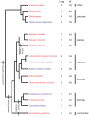
Figure 2. Phylogeny of species used, with our hypothesis of the lung reduction steps mapped on.
Red species were used for lung typification only, blue species for lung and vascular development only, and purple species for both. ‘Lung’ and ‘PA’ give lung type and pulmonary artery type (Fig. 4), respectively. Stem and family branches are based on Refs. –, ; the branches within families are based on Refs. , , .
Figure 3
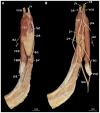
Figure 3. Example of annotation methods in Amira 5.4.5.
Dorsal side is up. V  =  ventricle. Scale bar represents 1 mm.
Figure 4
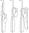
Figure 4. Schematic portrayal of pulmonary artery types and the lungs to which they connect.
Ventral views. PA1: pulmonary trunk that bifurcates into an ascending branch (connects with tracheal lung; tl) and a descending branch on the right (connects with the right lung; rl). PA2: single pulmonary artery, descending posteriorly and connecting to the right lung (rl); PA3: pulmonary trunk that bifurcates into two descending branches, each connecting to its respective lung (rl and ll); lat  =  left atrium; lpa  =  left pulmonary artery; pa  =  pulmonary artery; paa  =  anterior pulmonary artery; rat  =  right atrium; ptr  =  pulmonary trunk; rpa  =  right pulmonary artery; t  =  trachea; v  =  ventricle.
Figure 5
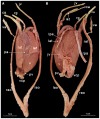
Figure 5. The Trimeresurus spp. Heart (ID PK07).
A Ventral view; B Dorsal view. az  =  azygos vein; ca  =  carotid artery; jv  =  jugular vein; lao  =  left aorta; lat  =  left atrium; lu  =  lung; paa  =  pulmonary artery anterior; pap  =  pulmonary artery posterior; pva  =  pulmonary vein anterior; pvp  =  pulmonary vein posterior; rao  =  right aorta; rat  =  right atrium; v  =  ventricle; va  =  vertebral artery; vca  =  vena cava anterior; vcp  =  vena cava posterior.
Figure 6
Figure 6. The Hydrophis elegans Heart (ID BS22).
A Ventral view; B Dorsal view. ca  =  carotid artery; jv  =  jugular vein; lao  =  left aorta; lat  =  left atrium; lu  =  lung; paa  =  pulmonary artery anterior; pap  =  pulmonary artery posterior; pva  =  pulmonary vein anterior; pvp  =  pulmonary vein posterior; rao  =  right aorta; rat  =  right atrium; v  =  ventricle; va  =  vertebral artery; vca  =  vena cava anterior; vcp  =  vena cava posterior.
Figure 7
Figure 7. The Bungarus candidus heart (ID BS24).
A Ventral view; B Dorsal view. The lung was turned laterally by 180 degrees for the purpose of these photographs, which has detached the pulmonary vein and vena cava posterior that normally run together. az  =  azygous artery; ca  =  carotid artery; jv  =  jugular vein; lao  =  left aorta; lat  =  left atrium; lu  =  lung; pa  =  pulmonary artery; pv  =  pulmonary vein; rao  =  right aorta; rat  =  right atrium; v  =  ventricle; va  =  vertebral artery; vca  =  vena cava anterior; vcp  =  vena cava posterior.
Figure 8
Figure 8. The Eunectus notaeus heart (ID BS8).
A ventral view; B dorsal view. az  =  azygos vein; ca  =  carotid artery; jv  =  jugular vein; lao  =  left aorta; lat  =  left atrium; pv  =  pulmonary vein; lpa  =  left pulmonary artery; rao  =  right aorta; rat  =  right atrium; rpa  =  right pulmonary artery; rg  =  ramus glandularis; v  =  ventricle; va  =  vertebral artery; vca  =  vena cava anterior; vcp  =  vena cava posterior.
Figure 9
Figure 9. Legend for 3D segmentations in Amira.
Figure 10
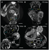
Figure 10. MicroCT transverse sections of embryos showing lung lumina.
Sections are of Pantherophis guttata guttata (A – D) and Elaphe obsoleta spiloides (E). The embryo's dorsal sides are up. Astral blue arrows denote right lung, dark yellow left lung; V  =  ventricle; OFT  =  outflow tract. Annotation done in Amira 5.4.5. Scale bar represents 1 mm.
Figure 11
Figure 11. MicroCT transverse sections of embryos showing lung lumina.
Sections are of Python curtus breitensteini. The embryo's dorsal sides are up. Astral blue arrows denote right lung, dark yellow left lung; V  =  ventricle; OFT  =  outflow tract. Annotation done in Amira 5.4.5. Scale bar represents 1 mm.
Figure 12
Figure 12. MicroCT transverse sections of embryos showing lung lumina.
Sections are of Causus rhombeatus (A – C) and Calloselasma rhodostoma (D, E). The embryo's dorsal sides are up. Astral blue arrows denote right lung, dark yellow left lung; V  =  ventricle. Annotation done in Amira 5.4.5. Scale bar represents 1 mm.
Figure 13
Figure 13. Overview of 3D reconstructions comparing similar developmental stages (Zehr 22–24
[40] ). Visualized species are Pantherophis guttata guttata (A), Python curtus breitensteini (B) and Causus rhombeatus (C). Reconstructions were made in Amira 5.4.5. Dashed lines denote outflow tract contours. Scale bars represent 1 mm.
Figure 14
Figure 14. Overview of 3D reconstructions comparing similar developmental stages (Zehr 25–28
[40] ). Visualized species are Pantherophis guttata guttata (A), Python curtus breitensteini (B) and Causus rhombeatus (C). Reconstructions were made in Amira 5.4.5. Dashed lines denote outflow tract contours. Scale bars represent 1 mm.
Figure 15
Figure 15. Overview of 3D reconstructions comparing similar developmental stages (Zehr 26–30
[40] ). Visualized species are Pantherophis guttata guttata (A), Python curtus breitensteini (B), Causus rhombeatus (C) and calloselasma rhodostoma (D). Reconstructions were made in Amira 5.4.5.Dashed lines denote outflow tract contours. Scale bars represent 1 mm.
Figure 16
Figure 16. Overview of 3D reconstructions comparing similar developmental stages (Zehr 32–33
[40] ). Visualized species are Pantherophis guttata guttata (A) and Python curtus breitensteini (B). Reconstructions were made in Amira 5.4.5. Dashed lines denote outflow tract contours. Scale bars represent 1 mm.
Figure 17
Figure 17. Overview of 3D reconstructions comparing similar developmental stages (Zehr 34–36
[40] ). Visualized species are Elaphe obsolete spiloides (A), and Calloselasma rhodostoma (B). Reconstructions were made in Amira 5.4.5. Scale bars represent 1 mm.

References

    1. Metzger RJ, Klein OD, Martin GR, Krasnow MA (2008) The branching programme of mouse lung development. Nature 453:745–750. - PMC - PubMed
    1. Cardoso WV, Lü J (2006) Regulation of early lung morphogenesis: questions, facts and controversies. Development 133:1611–1624. - PubMed
    1. Cardoso WV (2001) Molecular regulation of lung development. Annu Rev Physiol 63:471–494. - PubMed
    1. Lin CR, Kioussi C, O’Connell S, Briata P, Szeto D, et al. (1999) Pitx2 regulates lung asymmetry, cardiac positioning and pituitary and tooth morphogenesis. Nature 401:279–282. - PubMed
    1. Arora R, Metzger RJ, Papaioannou VE (2012) Multiple roles and interactions of Tbx4 and Tbx5 in development of the respiratory system. PLoS Genet 8:e1002866. - PMC - PubMed

Publication types

MeSH terms

LinkOut - more resources