. 2015 Feb 20;290(8):5105-5116.
doi: 10.1074/jbc.M114.599969. Epub 2015 Jan 2.

Subunit-specific regulation of N-methyl-D-aspartate (NMDA) receptor trafficking by SAP102 protein splice variants

Affiliations

Subunit-specific regulation of N-methyl-D-aspartate (NMDA) receptor trafficking by SAP102 protein splice variants

Zhe Wei et al. J Biol Chem. .

Abstract

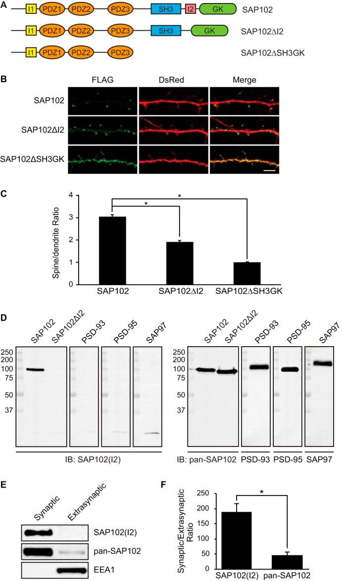
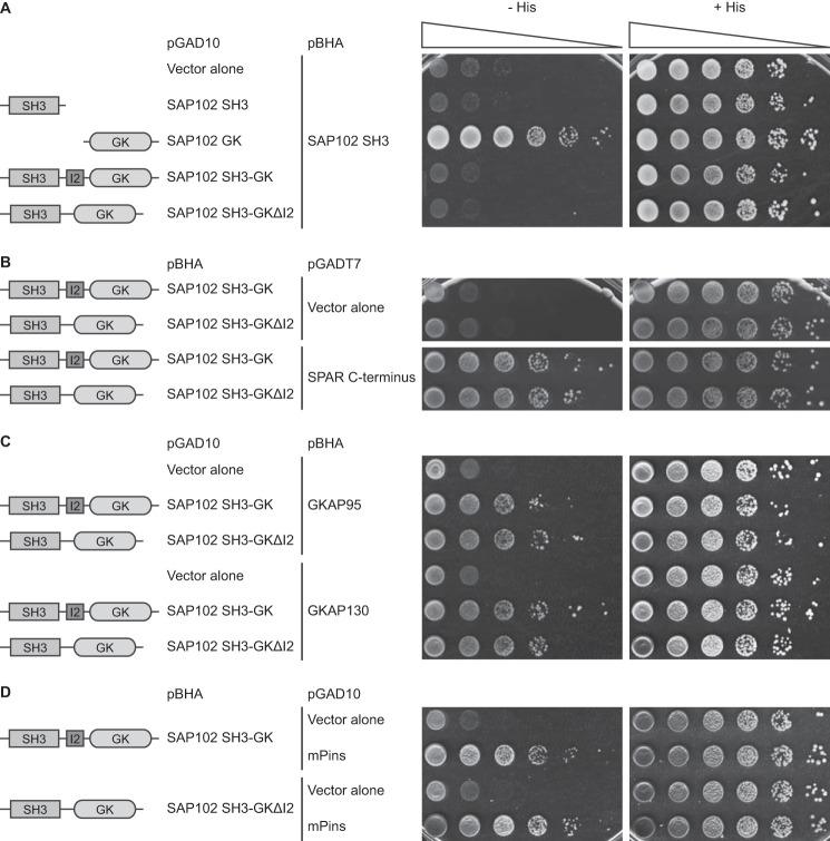
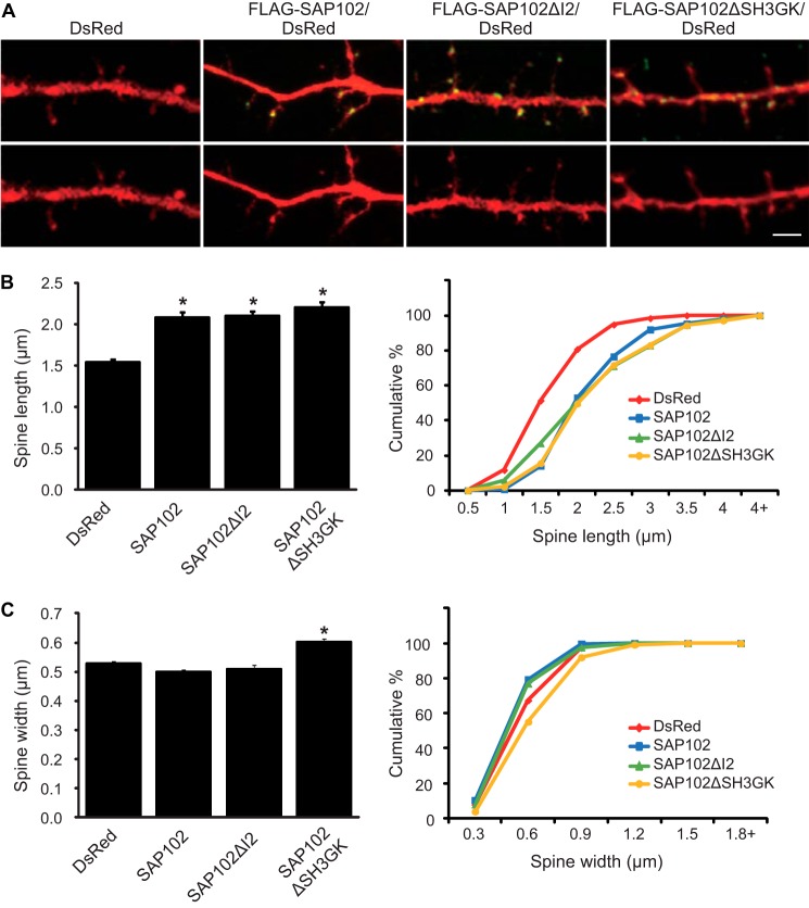
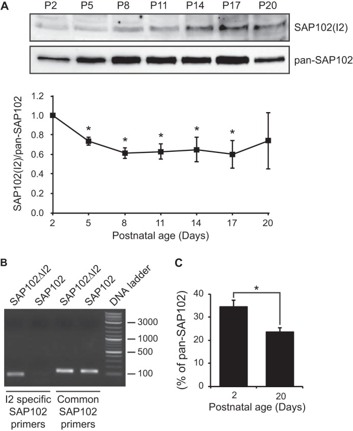
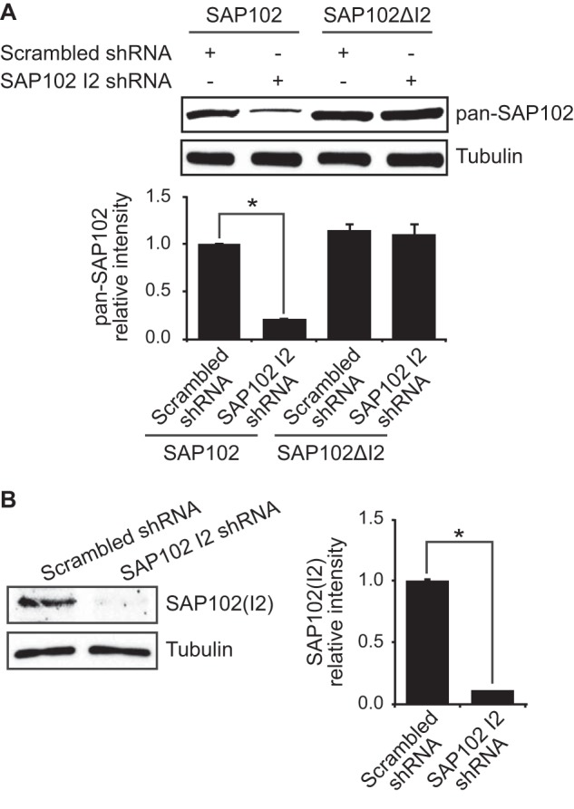
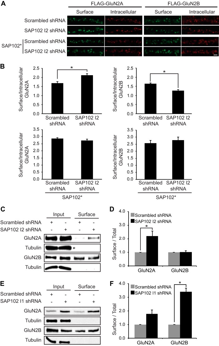
Synapse-associated protein 102 (SAP102) is a scaffolding protein abundantly expressed early in development that mediates glutamate receptor trafficking during synaptogenesis. Mutations in human SAP102 have been reported to cause intellectual disability, which is consistent with its important role during early postnatal development. SAP102 contains PDZ, SH3, and guanylate kinase (GK)-like domains, which mediate specific protein-protein interactions. SAP102 binds directly to N-methyl-D-aspartate receptors (NMDARs), anchors receptors at synapses, and facilitates transduction of NMDAR signals. Proper localization of SAP102 at the postsynaptic density is essential to these functions. However, how SAP102 is targeted to synapses is unclear. In the current study we find that synaptic localization of SAP102 is regulated by alternative splicing. The SAP102 splice variant that possesses a C-terminal insert (I2) between the SH3 and GK domains is highly enriched at dendritic spines. We also show that there is an intramolecular interaction between the SH3 and GK domains in SAP102 but that the I2 splicing does not influence SH3-GK interaction. Previously, we have shown that SAP102 expression promotes spine lengthening. We now find that the spine lengthening effect is independent of the C-terminal alternative splicing of SAP102. In addition, expression of I2-containing SAP102 isoforms is regulated developmentally. Knockdown of endogenous I2-containing SAP102 isoforms differentially affect NMDAR surface expression in a subunit-specific manner. These data shed new light on the role of SAP102 in the regulation of NMDAR trafficking.

Keywords: Glutamate Receptor; MAGUK; NMDA; PDZ Domain; Protein Splicing; Protein Targeting; SAP102; Scaffold Protein.


Figures

FIGURE 1.
FIGURE 1.
The I2 region is important for SAP102 clustering in spines. A, schematic domain structures of two naturally occurring splice variants of SAP102 (SAP102 and SAP102 ΔI2) and a C-terminal truncated form used in this study. SAP102 contains both I1 and I2 regions. SAP102ΔI2 contains a 14-aa deletion of the I2 region between the SH3 and GK domains (14). B, hippocampal neurons were co-transfected with FLAG-SAP102/DsRed, FLAG-SAP102ΔI2/DsRed, or FLAG-SAP102ΔSH3GK/DsRed. SAP102ΔI2 displayed reduced synaptic targeting. FLAG-SAP102 is enriched in spines. FLAG-SAP102ΔSH3GK is uniformly distributed throughout the dendrite and the spines. Scale bar, 5 μm. C, the mean fluorescence intensity of spines compared with that in adjacent dendrites. The spine/dendrite green fluorescence ratio of FLAG-SAP102, FLAG-SAP102ΔI2, and FLAG-SAP102ΔSH3GK is 3 ± 0.09, 1.9 ± 0.05, and 0.98 ± 0.03, respectively. n = 10–15 neurons from three transfections. *, p < 0.05, t test with Bonferroni's correction after ANOVA. D, HEK-293 cells were transfected with SAP102, SAP102ΔI2, PSD-93, PSD-95, or SAP97, and immunoblots (IB) of cell lysate were probed with pan-SAP102, SAP102 I2-specific, PSD-93, PSD-95, or SAP97 antibody. E, distribution of proteins in synaptic and extrasynaptic membrane fractions. Equal amounts of total proteins were loaded from each of the fractions and probed with I2-specific SAP102 antibody, pan-SAP102 antibody (detects all SAP102 splice variants), and the early endosomal marker EEA1 antibody. F, ratio of synaptic to extrasynaptic membrane fractions of SAP102(I2) and total SAP102. Data represent the means ± S.E. The experiment was repeated three times and quantified using ImageQuant LAS TL software. *, p < 0.05, Student's t test.
FIGURE 2.
FIGURE 2.
Deletion of the I2 region does not affect the intramolecular interaction between the SH3 and GK domains in SAP102 and the SPAR, GKAP, and mPins binding. Yeast were co-transformed with pBHA-SAP102 SH3 and pGAD10-SAP102 SH3, GK, SH3-GK or SH3-GKΔI2 (A), pGADT7-SPAR C terminus and pBHA-SAP102 SH3-GK or SH-GKΔI2 (B), pGAD10-SAP102 SH3-GK or SH-GKΔI2 and pBHA-GKAP95 or GKAP130 (C), and pGAD10-SAP102 SH3-GK or SH-GKΔI2 and pBHA-mPins (D), and growth was evaluated on appropriate yeast selection medium. Results shown are 10-fold serial dilutions of yeast cells.
FIGURE 3.
FIGURE 3.
SAP102 splice variants regulate spine morphology. A, primary hippocampal neurons (DIV12) were transfected with FLAG-SAP102/DsRed, FLAG-SAP102ΔI2/DsRed, FLAG-SAP102ΔSH3GK, or DsRed only. At DIV14, they were fixed and labeled with anti-FLAG antibody (green). Scale bar, 2 μm. B, dendritic spines were quantified by measuring DsRed signal using ImageJ software. Dendrites on neurons transfected with FLAG-SAP102/DsRed or FLAG-SAP102ΔI2/DsRed had longer protrusions than those transfected with FLAG-SAP102 ΔSH3GK/DsRed or DsRed only. C, cumulative frequency plots of spine length and spine width are shown. Data represent the means ± S.E. (n = 10 neurons per condition from 3 independent cultures; 20–30 spines/neuron; *, p < 0.05, t test with Bonferroni's correction after ANOVA).
FIGURE 4.
FIGURE 4.
SAP102 splice variants are developmentally regulated. A, whole rat brain lysate was collected at various ages. The P2 fraction was isolated and resolved by 10% SDS-PAGE. Samples were immunoblotted with either pan-SAP102 antibody or with I2-specific SAP102 antibody. The experiment was repeated three times and quantified using ImageQuant LAS TL software. A representative blot is shown in A. All ages were normalized to the intensity at P2, and a ratio of SAP102(I2) to total SAP102 was determined. Data represent the means ± S.E. (*, p < 0.05, one-sample t test). B, specificity of I2-specific and common SAP102 primers was confirmed by PCR with FLAG-SAP102ΔI2 and FLAG-SAP102. C, the mRNA levels of SAP102(I2) were measured by performing real-time PCR with cDNA from mouse cortex at postnatal day 2 and 20. Samples were run in triplicate (n = 3). Data represent the means ± S.E. (*, p < 0.05, Student's t test).
FIGURE 5.
FIGURE 5.
shRNA knockdown of endogenous I2-containing SAP102. A, HEK-293 cells were co-transfected with SAP102 or SAP102ΔI2 and scrambled shRNA or SAP102 I2 shRNA, and the immunoblots of cell lysate were probed with pan-SAP102 or tubulin antibody. Data represent the means ± S.E. (n = 3 independent experiments; *, p < 0.05, Student's t test). B, primary cortical neurons (DIV4) were infected with lentivirus containing SAP102 I2 shRNA or scrambled shRNA. The cell lysates were prepared at DIV14, resolved by SDS-PAGE, and immunoblotted with I2-specific SAP102 or tubulin antibody. Data represent the means ± S.E. (n = 3 independent experiments; *, p < 0.05, Student's t test).
FIGURE 6.
FIGURE 6.
Knock-down of I2-containing SAP102 increases surface expression of GluN2A. A, I2-containing SAP102 was knocked down in hippocampal cultures by the lentiviral induction of SAP102 I2 shRNA at DIV4, and FLAG-tagged GluN2A or GluN2B was transfected at DIV12. B, co-transfection of FLAG-tagged GluN2A or GluN2B with an shRNA-proofed Myc-tagged SAP102 variant (SAP102*). Surface staining was performed at DIV14 with anti-FLAG and Alexa 647 secondary antibodies (green), and after permeabilization, the intracellular pool was labeled with anti-FLAG and Alexa 568 secondary antibodies (red). Scale bar, 2 μm. B, quantification of the imaging experiments. Fluorescence intensities were measured using ImageJ software. Data represent the means ± S.E. (n = 27; n = 3 independent experiments); *, p < 0.05, Student's t test. C, biotinylated surface proteins from cortical neurons infected with lentivirus containing scrambled or SAP102 I2 shRNA were isolated, resolved by SDS-PAGE, and probed with GluN2A, GluN2B, or α-tubulin antibodies. D, quantification of the biotinylation experiments. The immunoreactive signals for surface GluN2A and GluN2B were normalized to total input and presented as a bar graph. n = 3 independent experiments. Data represent the mean ± S.E.; *, p < 0.05, Student's t test. E, biotinylated surface proteins from cortical neurons infected with lentivirus containing scrambled or SAP102 I1 shRNA were isolated, resolved by SDS-PAGE, and probed with GluN2A, GluN2B, or α-tubulin antibodies. F, quantification of the biotinylation experiments. The immune-reactive signals for surface GluN2A and GluN2B were normalized to total input and presented as a bar graph. n = 3 independent experiments. Data represent the means ± S.E.; *, p < 0.05, Student's t test.

References

    1. Elias G. M., Nicoll R. A. (2007) Synaptic trafficking of glutamate receptors by MAGUK scaffolding proteins. Trends Cell Biol. 17, 343–352 - PubMed
    1. El-Husseini A. E., Topinka J. R., Lehrer-Graiwer J. E., Firestein B. L., Craven S. E., Aoki C., Bredt D. S. (2000) Ion channel clustering by membrane-associated guanylate kinases. Differential regulation by N-terminal lipid and metal binding motifs. J. Biol. Chem. 275, 23904–23910 - PubMed
    1. Cuthbert P. C., Stanford L. E., Coba M. P., Ainge J. A., Fink A. E., Opazo P., Delgado J. Y., Komiyama N. H., O'Dell T. J., Grant S. G. (2007) Synapse-associated protein 102/dlgh3 couples the NMDA receptor to specific plasticity pathways and learning strategies. J. Neurosci. 27, 2673–2682 - PMC - PubMed
    1. Sans N., Petralia R. S., Wang Y. X., Blahos J., 2nd, Hell J. W., Wenthold R. J. (2000) A developmental change in NMDA receptor-associated proteins at hippocampal synapses. J. Neurosci. 20, 1260–1271 - PMC - PubMed
    1. Sans N., Prybylowski K., Petralia R. S., Chang K., Wang Y. X., Racca C., Vicini S., Wenthold R. J. (2003) NMDA receptor trafficking through an interaction between PDZ proteins and the exocyst complex. Nat. Cell Biol. 5, 520–530 - PubMed

Publication types

LinkOut - more resources