Review
. 2015 Mar;129(3):363-81.
doi: 10.1007/s00401-014-1379-7. Epub 2015 Jan 3.

Early etiology of Alzheimer's disease: tipping the balance toward autophagy or endosomal dysfunction?

Affiliations
Review

Early etiology of Alzheimer's disease: tipping the balance toward autophagy or endosomal dysfunction?

Aleksandar Peric et al. Acta Neuropathol. 2015 Mar.

Abstract

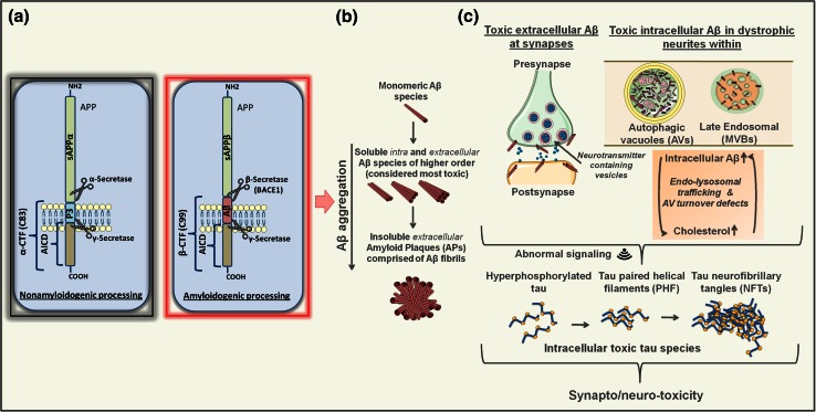
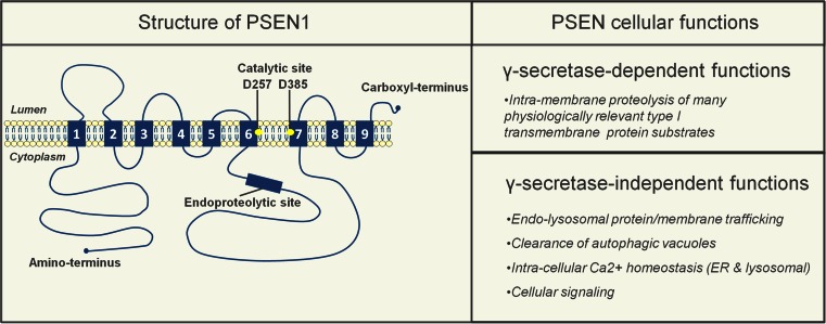
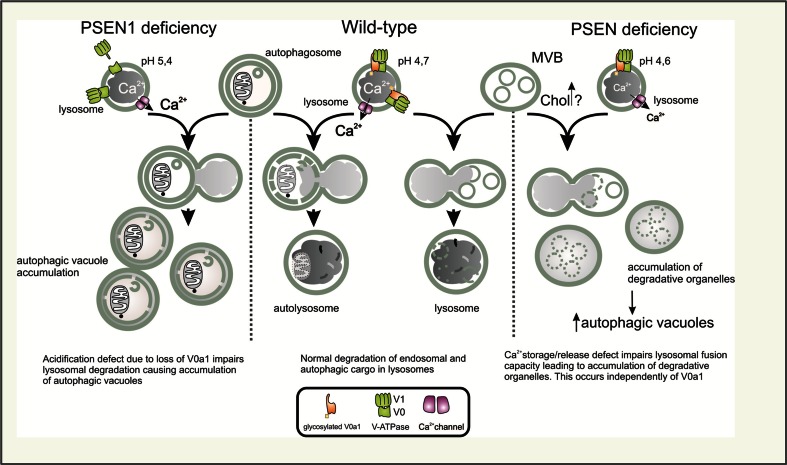
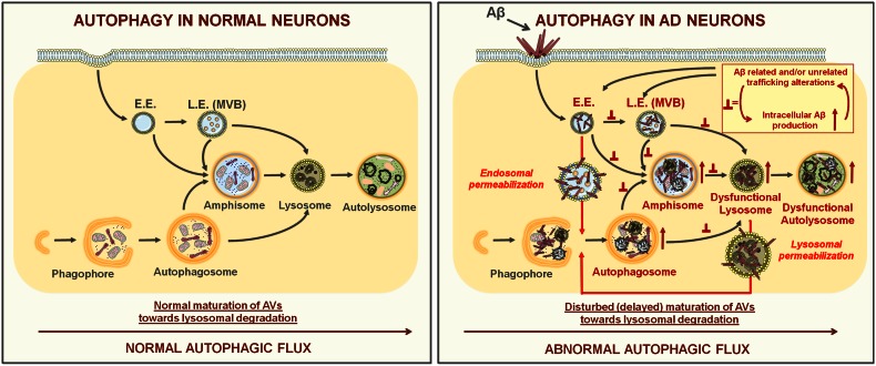
Alzheimer's disease (AD) is the most common form of dementia in the elderly. This brain neuropathology is characterized by a progressive synaptic dysfunction and neuronal loss, which lead to decline in memory and other cognitive functions. Histopathologically, AD manifests via synaptic abnormalities, neuronal degeneration as well as the deposition of extracellular amyloid plaques and intraneuronal neurofibrillary tangles. While the exact pathogenic contribution of these two AD hallmarks and their abundant constituents [aggregation-prone amyloid β (Aβ) peptide species and hyperphosphorylated tau protein, respectively] remain debated, a growing body of evidence suggests that their development may be paralleled or even preceded by the alterations/dysfunctions in the endolysosomal and the autophagic system. In AD-affected neurons, abnormalities in these cellular pathways are readily observed already at early stages of disease development, and even though many studies agree that defective lysosomal degradation may relate to or even underlie some of these deficits, specific upstream molecular defects are still deliberated. In this review we summarize various pathogenic events that may lead to these cellular abnormalities, in light of our current understanding of molecular mechanisms that govern AD progression. In addition, we also highlight the increasing evidence supporting mutual functional dependence of the endolysosomal trafficking and autophagy, in particular focusing on those molecules and processes which may be of significance to AD.


Figures

Fig. 1
Fig. 1
Amyloidogenic vs. nonamyloidogenic APP processing, Aβ aggregation and intracellular vs. extracellular Aβ toxicity. a Non-amyloidogenic processing of APP (left) requires the dual proteolysis, first by members of the ADAM (a disintegrin and metalloprotease domain-containing) protein family (mainly, ADAM10 and ADAM17, also called α-secretases) followed by γ-secretase, resulting in release of soluble sAPPα ectodomain, a non-amyloidogenic p3 fragment and APP intracellular domain (AICD). In the amyloidogenic pathway (right), APP is first cleaved by β-secretase BACE1 releasing the sAPPβ ectodomain, followed by γ-secretase processing of the remaining β-CTF giving rise to AICD and Aβ peptide species of slightly different lengths. b Monomeric Aβ species, particularly Aβ42 and Aβ43, have a tendency to aggregate and form structures of higher order. These include toxic soluble Aβ dimers, trimers, oligomers and protofibrils, found both inside and outside of the cells/neurons, as well as more inert insoluble amyloid fibrils, which comprise extracellular APs. c In AD, in addition to their direct effects on synaptic transmission/integrity via binding to synaptic membranes/receptors, toxic Aβ species may also accumulate within dystrophic neurites and aberrant synaptic regions in intracellular compartments, including late endosomal multivesicular bodies (MVB) and autophagic vacuoles (AVs) [144, 172]. Indicated as well is the hypothesized self-propelling exacerbating influence of excessive Aβ/cholesterol accumulation, disturbed endolysosomal trafficking regulation and/or defective turnover of autophagic vacuoles (AV) in this context. All together, this could lead to aberrant cellular signaling that in turn may induce and propagate excessive tau phosphorylation, accumulation of toxic tau species and related synapto/neurotoxic effects
Fig. 2
Fig. 2
γ-secretase-dependent and -independent functions of presenilins (PSENs). PSEN1 (and likely PSEN2) has nine transmembrane domains (TMDs) [139], with two aspartate residues (D257/D385; yellow circles) in TMD 6 and 7 forming the catalytic core [138]. Full-length PSEN1 is endoproteolyzed in early secretory compartments resulting in stable PSEN1-NTF and -CTF heterodimers [138]. The catalytic role of PSENs, as part of the γ-secretase complex, is associated with the processing of around 100 currently known substrates [60]. PSENs, however, also have γ-secretase-independent functions, including roles in endolysosomal protein/membrane trafficking and clearance of autophagic vacuoles [37, 69, 116, 164], intracellular Ca2+ homeostasis (ER [98, 153] and lysosomal [24, 96]) and cellular signaling [6, 7, 33, 116]
Fig. 3
Fig. 3
Presenilins (PSENs) and lysosomal degradation. Impaired lysosomal degradation observed in PSEN-deficient cells is attributed to either a failure in lysosomal acidification (left) or disturbed lysosomal calcium release/storage (right). Left a defect in lysosomal acidification is here primarily caused by the failure of the V0a1 subunit of the V-ATPase (proton pump) to become N-glycosylated, resulting in a dysfunctional proton pump, higher pH and decreased lysosomal degradative capacity. This in turn is claimed to underlie the accumulation of autophagic vacuoles (AVs) [69]. Alternatively (right), a deficit in lysosomal calcium storage/release affects the fusion of degradation-prone vesicles (late endosomes (MVBs) and AVs) with lysosomes [24, 37], thus compromising their clearance. Here, PSEN-dependent lysosomal Ca2+ defects could relate to alterations in endosomal trafficking homeostasis (endosomal recycling and normal endosomal maturation), which may lead to a buildup of lipids like cholesterol (Chol) and/or mislocalization of relevant transporters and channels, all of which could underlie the observed deficits (see the main text for clarifications). The middle panel depicts undisturbed fusion/degradation with/in lysosomes in cells with normal levels of PSENs
Fig. 4
Fig. 4
Stepwise autophagy progression in normal and AD-affected neurons. Left different steps in autophagosome formation and maturation under normal (physiological) conditions, starting from phagophore expansion. Before fusing with lysosomes, double-membraned autophagosomes can also merge with early (E.E) and late endosomes (L.E./MVBs) giving rise to amphisomes. Right in AD-related processes, normal autophagic flux is compromised. This results in a pile up of autophagic vacuoles (autophagosomes, amphisomes, autolysosomes) due to disturbed trafficking, fusion with and/or degradation processes within dysfunctional lysosomes. These changes are particularly pronounced in dystrophic neurites, where they likely contribute to synapto-/neurotoxicity (see also Fig. 1c). Here, intracellular toxic Aβ species may work together with dysfunctional endosomal sorting/trafficking mechanisms in aggravating these degradative abnormalities. Inset in the right panel depicts this hypothesized self-propelling endosomal dysfunction, while the arrows from it point toward the likely sites where consequent disturbances in membrane flow “roadblocks” may occur. These pathogenic processes may eventually compromise the impermeability of endolysosomal compartments. Because autophagy can directly target toxic Aβ species [62] as well as injured lysosomes [80], we hypothesize that in the AD context both physically injured endosomes/lysosomes and toxic Aβ species released into cytosol may become its targets. Here, initially autophagy may be protective, but as the disease develops and the toxic burden exceeds cellular reparative capacity, neuronal death may follow

References

    1. Almeida CG, Takahashi RH, Gouras GK. Beta-amyloid accumulation impairs multivesicular body sorting by inhibiting the ubiquitin-proteasome system. J Neurosci. 2006;26(16):4277–4288. - PMC - PubMed
    1. Alonso AC, Grundke-Iqbal I, Iqbal K. Alzheimer’s disease hyperphosphorylated tau sequesters normal tau into tangles of filaments and disassembles microtubules. Nat Med. 1996;2(7):783–787. - PubMed
    1. Annaert WG, Esselens C, Baert V, Boeve C, Snellings G, Cupers P, Craessaerts K, De Strooper B. Interaction with telencephalin and the amyloid precursor protein predicts a ring structure for presenilins. Neuron. 2001;32(4):579–589. - PubMed
    1. Aqul A, Liu B, Ramirez CM, Pieper AA, Estill SJ, Burns DK, Repa JJ, Turley SD, Dietschy JM. Unesterified cholesterol accumulation in late endosomes/lysosomes causes neurodegeneration and is prevented by driving cholesterol export from this compartment. J Neurosci. 2011;31(25):9404–9413. - PMC - PubMed
    1. Arriagada PV, Growdon JH, Hedley-Whyte ET, Hyman BT. Neurofibrillary tangles but not senile plaques parallel duration and severity of Alzheimer’s disease. Neurology. 1992;42(3 Pt 1):631–639. - PubMed

Publication types