

Alcohol Withdrawal-Induced Seizure Susceptibility is Associated with an Upregulation of CaV1.3 Channels in the Rat Inferior Colliculus
- PMID: 25556199
- PMCID: PMC4458366
- DOI: 10.1093/ijnp/pyu123
Alcohol Withdrawal-Induced Seizure Susceptibility is Associated with an Upregulation of CaV1.3 Channels in the Rat Inferior Colliculus
Abstract
Background: We previously reported increased current density through L-type voltage-gated Ca(2+) (CaV1) channels in inferior colliculus (IC) neurons during alcohol withdrawal. However, the molecular correlate of this increased CaV1 current is currently unknown.
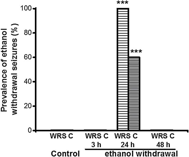
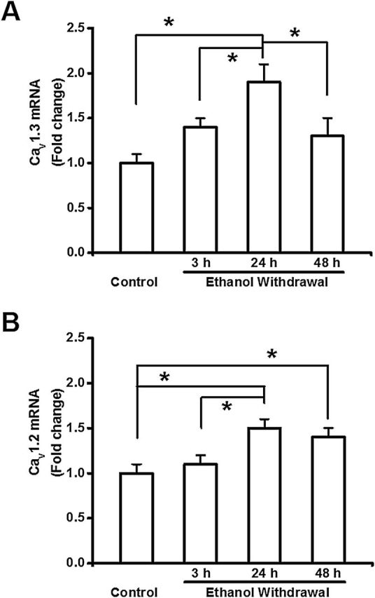
Methods: Rats received three daily doses of ethanol every 8 hours for 4 consecutive days; control rats received vehicle. The IC was dissected at various time intervals following alcohol withdrawal, and the mRNA and protein levels of the CaV1.3 and CaV1.2 α1 subunits were measured. In separate experiments, rats were tested for their susceptibility to alcohol withdrawal-induced seizures (AWS) 3, 24, and 48 hours after alcohol withdrawal.
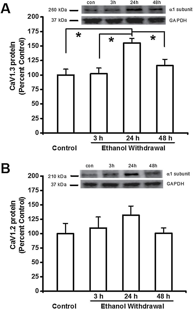
Results: In the alcohol-treated group, AWS were observed 24 hours after withdrawal; no seizures were observed at 3 or 48 hours. No seizures were observed at any time in the control-treated rats. Compared to control-treated rats, the mRNA level of the CaV1.3 α1 subunit was increased 1.4-fold, 1.9-fold, and 1.3-fold at 3, 24, and 48 hours, respectively. In contrast, the mRNA level of the CaV1.2 α1 subunit increased 1.5-fold and 1.4-fold at 24 and 48 hours, respectively. At 24 hours, Western blot analyses revealed that the levels of the CaV1.3 and CaV1.2 α1 subunits increased by 52% and 32%, respectively, 24 hours after alcohol withdrawal. In contrast, the CaV1.2 and CaV1.3 α1 subunits were not altered at either 3 or 48 hours during alcohol withdrawal.
Conclusions: Expression of the CaV1.3 α1 subunit increased in parallel with AWS development, suggesting that altered L-type CaV1.3 channel expression is an important feature of AWS pathogenesis.
Keywords: Cacna1c mRNA; Cacna1d mRNA; Cav1.2 α1 subunit; Cav1.3 α1 subunit; alcohol withdrawal seizures.
© The Author 2015. Published by Oxford University Press on behalf of CINP.
Figures
References
-
- Chakravarty DN, Faingold CL. (1998). Comparison of neuronal response patterns in the external and central nuclei of inferior colliculus during ethanol administration and ethanol withdrawal. Brain Res 783:102–108. - PubMed
-
- Eckardt MJ, Campbell GA, Marietta CA, Majchrowicz E, Wixon HN, Weight FF. (1986). Cerebral 2-deoxyglucose uptake in rats during ethanol withdrawal and post-withdrawal. Brain Res 366:1–9. - PubMed
-
- Evans MS, Li Y, Faingold CL. (2000). Inferior colliculus intracellular response abnormalities in vitro associated with susceptibility to ethanol withdrawal seizures. Alcohol Clin Exp Res 24:1180–1186. - PubMed
Publication types
MeSH terms
Substances
Grants and funding
LinkOut - more resources
Full Text Sources
Other Literature Sources
Miscellaneous
