

Review
doi: 10.5732/cjc.014.10273.
Pathobiology of ovarian carcinomas
Affiliations
- PMID: 25556618
- PMCID: PMC4302089
- DOI: 10.5732/cjc.014.10273
Item in Clipboard
Review
Pathobiology of ovarian carcinomas
Chin J Cancer.
2015 Jan.
Abstract
Ovarian tumors comprise a heterogeneous group of lesions, displaying distinct tumor pathology and oncogenic potentiel. These tumors are subdivided into three main categories: epithelial, germ cell, and sex-cord stromal tumors. We report herein the newly described molecular abnormalities in epithelial ovarian cancers (carcinomas). Immunohistochemistry and molecular testing help pathologists to decipher the significant heterogeneity of this disease. Our better understanding of the molecular basis of ovarian carcinomas represents the first step in the development of targeted therapies in the near future.
Figures
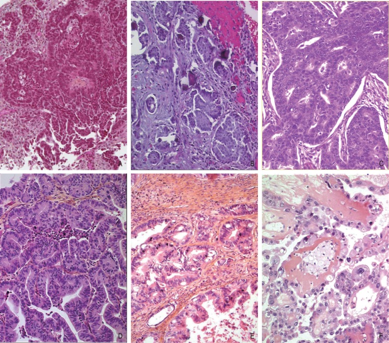
A, high-grade serous carcinoma is composed of a solid mass of cells with slit-like spaces. Nuclei are large, hyperchromatic, and pleomorphic with high mitotic activity (>12 mitoses per 10 high-power fields) (HES ×100). B, low-grade serous carcinoma is composed of micro- or macropapillae lined by uniform small cells with limited nuclear pleomorphisms and low mitotic activity (<12 mitoses per 10 high-power fields), associated with psammoma bodies (HES ×100). C, endometrioid carcinoma is arranged in a cribriform pattern lined by cuboidal cells with eosinophilic cytoplasm and moderate nuclear atypia (HES ×200). D, the expansile pattern of mucinous carcinoma shows proliferation of complex glands arranged back to back with limited intervening stroma, exhibiting moderate cytologic atypia (HES ×200). E, destructive pattern of mucinous carcinoma shows proliferation of irregular glands, nests, and cells with malignant cytology, infiltrating ovarian stroma (HES ×200). F, clear cell carcinoma shows papillae lined by cuboidal, hobnail cells with clear or eosinophilic cytoplasm and atypical nuclei. The papillae are round and small with a dense hyaline basement membrane material forming the core of the papillary stalk (HES ×200).
References
-
- Kurman RJ, Carcangiu ML, Herrington CS, et al. WHO classification of tumours of female reproductive organs. Lyon: IARC Press; 2014.
-
- Yanick J, Ries LG, Yates JW. Ovarian cancer in the elderly: an analysis of surveillance. J Obstet Gynecol. 1986;154:639–647. - PubMed
-
- Lauchlan SC. Metaplasia and neoplasias of the Müllerian epithelium. Histopathology. 1884;8:543–557. - PubMed
-
- Fathalla MF. Incessant ovulation—a factor in ovarian neoplasia. Lancet. 1971;2:163. - PubMed
-
- Prat J. New insights into ovarian cancer pathology. Ann Oncol. 2012;23(Suppl 10):111–117. - PubMed
Publication types
MeSH terms
LinkOut - more resources
Full Text Sources
Other Literature Sources
Medical