

Review
doi: 10.1113/jphysiol.2014.272724.
Epub 2014 May 6.
Glutamate receptor pores
Affiliations
- PMID: 25556787
- PMCID: PMC4293053
- DOI: 10.1113/jphysiol.2014.272724
Item in Clipboard
Review
Glutamate receptor pores
J Physiol.
.
Abstract
Glutamate receptors are ligand-gated ion channels that mediate fast excitatory synaptic transmission throughout the central nervous system. Functional receptors are homo- or heteromeric tetramers with each subunit contributing a re-entrant pore loop that dips into the membrane from the cytoplasmic side. The pore loops form a narrow constriction near their apex with a wide vestibule toward the cytoplasm and an aqueous central cavity facing the extracellular solution. This article focuses on the pore region, reviewing how structural differences among glutamate receptor subtypes determine their distinct functional properties.
© 2014 The Authors. The Journal of Physiology © 2014 The Physiological Society.
Figures
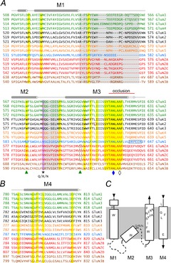
A, alignment of the M1–M3 segment for the four AMPA receptor subunits (green, UniProt numbers: P19490–19493), three low affinity KA receptor subunits capable of forming homomeric channels (black, P22756, P42260, P42264), two high affinity KA receptor subunits that do not form functional homomers (orange, Q01812, Q63273), the GluN1 subunit essential for all NMDA receptors (blue, P35439), four GluN2 subunits (red, Q00959–009561, Q62645) and two GluN3 subunits (brown, Q9R1M7, Q8VHN2). All sequences are from rat. Highly conserved residues are highlighted in yellow. Sequence that was not resolved in the GluA2 crystal structure is highlighted in grey. The asterisk indicates the Q/R/N site. GluA2, GluK1 and 2 are shown in the edited (R) form (red boxes). Green triangles indicate the M3 S/L site of GluN2 and the conserved M2 Trp of GluN1 that interact (green boxes). The GluN1 DRPEER motif is boxed in black. The conserved Asn (N5) and Lurcher site Ala (A8) in the SYTANLAAF motif are indicated by a filled blue diamond and open black diamond, respectively. Residue numbers in A and B are for the mature proteins after signal sequence removal. Secondary structure is shown as cylinders (α-helices) and lines (random coil loops) above the alignment. The red line above M3 indicates the occlusion at the inner helix bundle crossing in the GluA2 crystal structure. B, alignment of the M4 helix. C, diagram of TMD topology displaying the approximate position of the conserved residues highlighted in yellow in A and B. Dashed lines indicate segments that were not resolved in the GluA2 crystal structure.
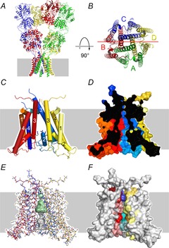
A, X-ray crystal structure of the homomeric GluA2 AMPA receptor (PDB 3KG2) from Sobolevsky et al. (2009). Grey background indicates the approximate position of the membrane. B, view down the pore axis from the extracellular membrane surface. The A/C (green/blue) and B/D (red/yellow) subunits are labelled. The horizontal red line indicates the plane of section through the TMD shown in side view in panel D. C–F, homology model of the rat GluK2(R) TMD from Lopez et al. (2013) based on the GluA2 crystal structure. For clarity, the A (green) subunit has been removed to provide a view into the pore. All four panels are shown in the same orientation. C, M1, M3 and M4 transmembrane helices and the M2 pore-loop helix shown as numbered cylinders. D, surface rendering sectioned along the axis of the pore as indicated in B. E, the pore cavity rendered as a surface generated by the program Hollow (Ho & Gruswitz, 2008). The central cavity is shaded green. The narrow constriction and cytoplasmic vestibule in grey are more speculative because residues in this region were disordered in the GluA2 crystal structure and were modelled without a template. F, coloured residues on this surface rendering are homologous to AMPA receptor locations that can be modified by extracellular or intracellular MTS reagents in the presence of agonist (Kuner et al. ; Sobolevsky et al. 2003). Darker colouration near the extracellular surface indicates residues within the bundle crossing occlusion zone.
References
-
- Antonov SM, Gmiro VE. Johnson JW. Binding sites for permeant ions in the channel of NMDA receptors and their effects on channel block. Nat Neurosci. 1998;1:451–461. - PubMed
-
- Beck C, Wollmuth LP, Seeburg PH, Sakmann B. Kuner T. NMDAR channel segments forming the extracellular vestibule inferred from the accessibility of substituted cysteines. Neuron. 1999;22:559–570. - PubMed
-
- Bernard A, Ferhat L, Dessi F, Charton G, Represa A, Ben-Ari Y. Khrestchatisky M. Q/R editing of the rat GluR5 and GluR6 kainate receptors in vivo and in vitro: evidence for independent developmental, pathological and cellular regulation. Eur J Neurosci. 1999;11:604–616. - PubMed
-
- Blanke ML. VanDongen AM. The NR1 M3 domain mediates allosteric coupling in the N-methyl-d-aspartate receptor. Mol Pharmacol. 2008;74:454–465. - PubMed
Publication types
MeSH terms
Substances
Grants and funding
LinkOut - more resources
Full Text Sources
Other Literature Sources
Molecular Biology Databases
Miscellaneous