. 2015 Jan 5:6:5904.
doi: 10.1038/ncomms6904.

Optical focusing deep inside dynamic scattering media with near-infrared time-reversed ultrasonically encoded (TRUE) light

Affiliations

Optical focusing deep inside dynamic scattering media with near-infrared time-reversed ultrasonically encoded (TRUE) light

Yan Liu et al. Nat Commun. .

Abstract

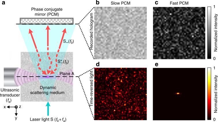
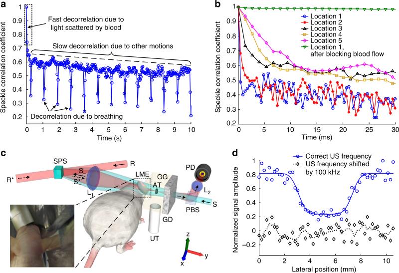
Focusing light deep inside living tissue has not been achieved despite its promise to play a central role in biomedical imaging, optical manipulation and therapy. To address this challenge, internal-guide-star-based wavefront engineering techniques--for example, time-reversed ultrasonically encoded (TRUE) optical focusing--were developed. The speeds of these techniques, however, were limited to no greater than 1 Hz, preventing them from in vivo applications. Here we improve the speed of optical focusing deep inside scattering media by two orders of magnitude, and focus diffuse light inside a dynamic scattering medium having a speckle correlation time as short as 5.6 ms, typical of living tissue. By imaging a target, we demonstrate the first focusing of diffuse light inside a dynamic scattering medium containing living tissue. Since the achieved focusing speed approaches the tissue decorrelation rate, this work is an important step towards in vivo deep tissue noninvasive optical imaging, optogenetics and photodynamic therapy.


Figures

Figure 1
Figure 1. The influence of phase conjugation speed on the quality of TRUE optical focusing
(a) Illustration of the TRUE focusing concept. Laser light S with a frequency of f0 + fa illuminates a scattering medium and a portion of the diffuse light traversing the acoustic focus is frequency-downshifted to f0 (the frequency of the acoustic wave is fa). A PCM records the wavefront of these ultrasonically modulated light S (f0) in a hologram and then phase-conjugates the light back to the ultrasonic focus, thereby forming a focus. Dashed arrows indicate time-reversed light. Plane A denotes the x–z plane intersecting the acoustic axis. (b,c) Simulated recorded holograms in a slow (b) and fast (c) PCM. Hologram blurring is clearly visible in b as a reduction in speckle contrast. (d,e) Simulated light intensity distribution on plane A by reading the hologram recorded in the slow (d) and fast (e) PCM. No focusing can be observed in d. All the images in b–e were normalized by their own maximum values. S, sample light; S , frequency-downshifted sample light (signal light); S, time-reversed signal light; TRUE, time-reversed ultrasonically encoded.
Figure 2
Figure 2. TRUE optical focusing inside a dynamic scattering medium containing a tissue-mimicking phantom
(a) Schematic of the set-up for characterizing the relationship between the speckle correlation time (τc) and the phantom movement speed. The front surface of the crystal and the object plane of the objective were mirrored planes. (b) The speckle correlation coefficient as a function of time when the phantom was moved at 0.010 mm s−1. τc = 60 ms was determined for this speed. (c) The relationship between the speckle correlation time and the phantom movement speed. Error bar shows the s.e. of τc measured when light illuminated three different locations on the intralipid–gelatin phantom. (d) Schematic of the set-up for evaluating the quality of the time-reversed light at various sample decorrelation rates. (e–g) The time-reversed light pattern when the intralipid–gelatin phantom was static (τc>300 s, e), moved at 0.100 mm s−1 (τc = 5.6 ms, f) and at 0.200 mm s 1 (τc = 2.8 ms, g). (h) No time-reversed light was observed when the frequency of S was shifted by 100 kHz (τc = 0.01 ms). All the images in e–h were normalized by their own maximum values. (i) Schematic of the set-up for imaging an absorptive target with TRUE light. The target was scanned along the x direction. (j) One-dimensional images of the target acquired under different conditions. The circles, squares and diamonds denote experimental data. The solid and dashed lines denote curve fitting of the experimental data. The dotted line denotes the four-point moving average of the experimental data. AT, absorptive target; GD, ground glass diffuser; GG, gelatin gel; IP, intralipid–gelatin phantom; L, lens; LT, lens tube; M, mirror; Obj, Objective; PBS, polarizing beamsplitter; PD, photodiode; R, reference beam; R*, reading beam, phase conjugate to R; S, sample light; S , frequency-downshifted sample light (signal light); S, time-reversed signal light; SPS, Sn2P2S6:Te 1% photorefractive crystal; TRUE, time-reversed ultrasonically encoded; US, ultrasound; UT, ultrasonic transducer. Scale bar, 1 mm.
Figure 3
Figure 3. TRUE optical focusing inside a dynamic scattering medium containing a living-mouse ear
(a) The speckle correlation coefficient as a function of time for a living-mouse ear. Three speckle decorrelation characteristics were identified. (b) The speckle correlation curves measured at five locations on the mouse ear. The speckle correlation time (τc) determined from the curves ranged from less than 0.44–10 ms. When the blood flow was blocked, τc became much larger. (c) Schematic of the set-up for imaging an absorptive target placed between a living-mouse ear and a diffuser. The target was scanned along the x direction. Inset: a photo showing the right ear of a mouse used as a dynamic scattering medium. The left ear was bent downwards to avoid blocking the light. Aluminium foil tapes were used to block the light that did not pass through the right ear. (d) 1D images of the absorptive target. The circles and diamonds denote experimental data. The solid line denotes curve fitting of the experimental data. The dotted line denotes the four-point moving average of the experimental data. AT, absorptive target; GD, ground glass diffuser; GG, gelatin gel; L, lens; LME, living-mouse ear; PBS, polarizing beamsplitter; PD, photodiode; R, reference beam; R*, reading beam, phase conjugate to R; S, sample light; S, frequency-downshifted sample light (signal light); S, time-reversed signal light; SPS, Sn2P2S6:Te 1% photorefractive crystal; TRUE, time-reversed ultrasonically encoded; US, ultrasound; UT, ultrasonic transducer.

Similar articles

Cited by

  • Rapid wide-field imaging through scattering media by digital holographic wavefront correction.
    Li R, Peng T, Zhou M, Yu X, Gao P, Min J, Yang Y, Lei M, Yao B, Zhang C, Ye T. Li R, et al. Appl Opt. 2019 Apr 10;58(11):2845-2853. doi: 10.1364/AO.58.002845. Appl Opt. 2019. PMID: 31044887 Free PMC article.
  • Focusing light into scattering media with ultrasound-induced field perturbation.
    Cheng Z, Wang LV. Cheng Z, et al. Light Sci Appl. 2021 Aug 2;10(1):159. doi: 10.1038/s41377-021-00605-7. Light Sci Appl. 2021. PMID: 34341328 Free PMC article.
  • Neurophotonic tools for microscopic measurements and manipulation: status report.
    Abdelfattah AS, Ahuja S, Akkin T, Allu SR, Brake J, Boas DA, Buckley EM, Campbell RE, Chen AI, Cheng X, Čižmár T, Costantini I, De Vittorio M, Devor A, Doran PR, El Khatib M, Emiliani V, Fomin-Thunemann N, Fainman Y, Fernandez-Alfonso T, Ferri CGL, Gilad A, Han X, Harris A, Hillman EMC, Hochgeschwender U, Holt MG, Ji N, Kılıç K, Lake EMR, Li L, Li T, Mächler P, Miller EW, Mesquita RC, Nadella KMNS, Nägerl UV, Nasu Y, Nimmerjahn A, Ondráčková P, Pavone FS, Perez Campos C, Peterka DS, Pisano F, Pisanello F, Puppo F, Sabatini BL, Sadegh S, Sakadzic S, Shoham S, Shroff SN, Silver RA, Sims RR, Smith SL, Srinivasan VJ, Thunemann M, Tian L, Tian L, Troxler T, Valera A, Vaziri A, Vinogradov SA, Vitale F, Wang LV, Uhlířová H, Xu C, Yang C, Yang MH, Yellen G, Yizhar O, Zhao Y. Abdelfattah AS, et al. Neurophotonics. 2022 Jan;9(Suppl 1):013001. doi: 10.1117/1.NPh.9.S1.013001. Epub 2022 Apr 27. Neurophotonics. 2022. PMID: 35493335 Free PMC article.
  • Ultrasound-modulated optical glucose sensing using a 1645 nm laser.
    Park EY, Baik J, Kim H, Park SM, Kim C. Park EY, et al. Sci Rep. 2020 Aug 7;10(1):13361. doi: 10.1038/s41598-020-70305-6. Sci Rep. 2020. PMID: 32770091 Free PMC article.
  • Single-shot time-reversed optical focusing into and through scattering media.
    Cheng Z, Yang J, Wang LV. Cheng Z, et al. ACS Photonics. 2020 Oct 21;7(10):2871-2877. doi: 10.1021/acsphotonics.0c01154. Epub 2020 Sep 18. ACS Photonics. 2020. PMID: 34337103 Free PMC article.

References

    1. Denk W, Strickler J, Webb W. Two-photon laser scanning fluorescence microscopy. Science. 1990;248:73–76. - PubMed
    1. Pawley J. Handbook of Biological Confocal Microscopy. Springer; 2010.
    1. Huang D, et al. Optical coherence tomography. Science. 1991;254:1178–1181. - PMC - PubMed
    1. Grier DG. A revolution in optical manipulation. Nature. 2003;424:810–816. - PubMed
    1. Boyden ES, Zhang F, Bamberg E, Nagel G, Deisseroth K. Millisecond-timescale, genetically targeted optical control of neural activity. Nat. Neurosci. 2005;8:1263–1268. - PubMed

Publication types