. 2015 Jan 15;160(1-2):191-203.
doi: 10.1016/j.cell.2014.11.041. Epub 2014 Dec 31.

Low affinity binding site clusters confer hox specificity and regulatory robustness

Affiliations

Low affinity binding site clusters confer hox specificity and regulatory robustness

Justin Crocker et al. Cell. .

Abstract

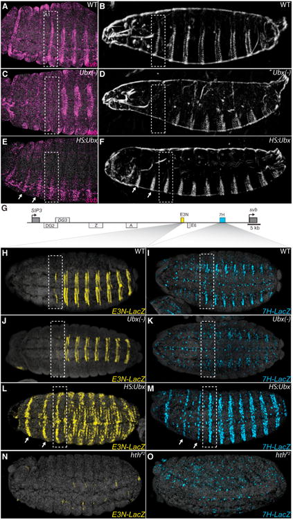
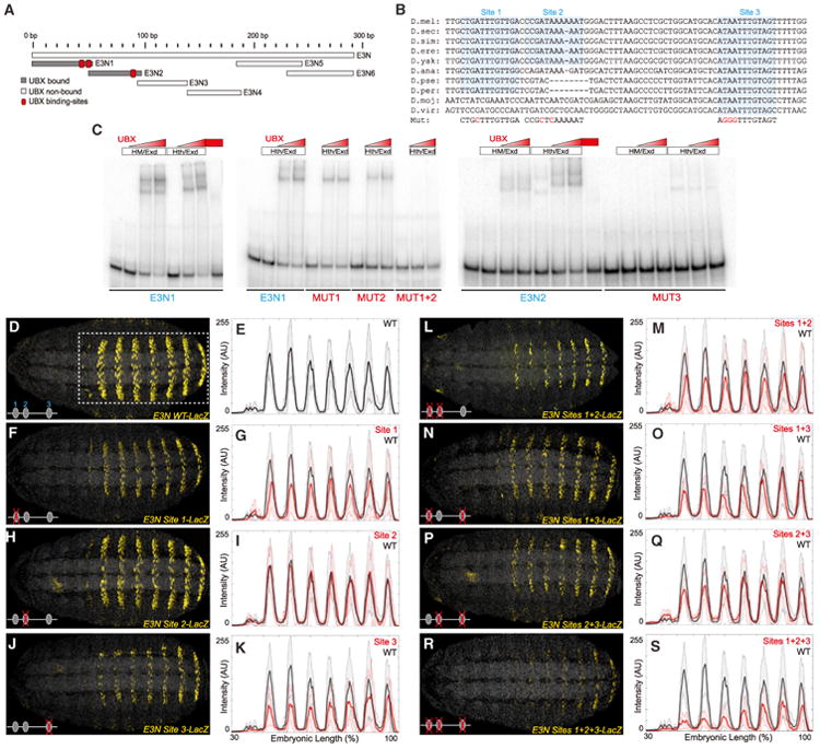
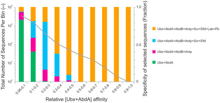
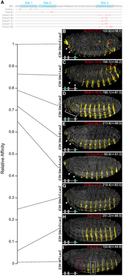
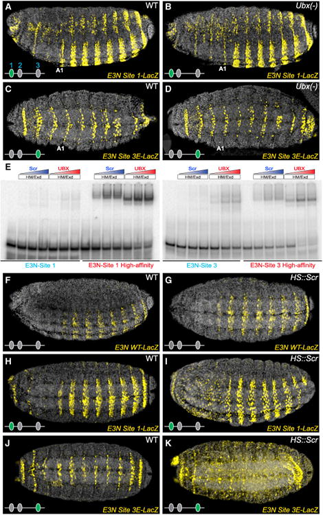
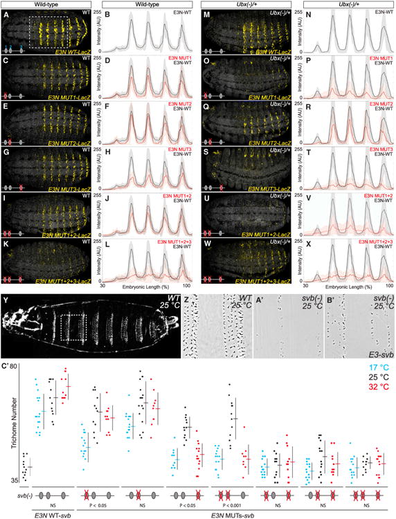
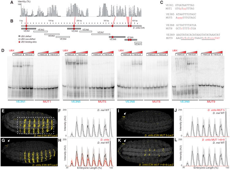
In animals, Hox transcription factors define regional identity in distinct anatomical domains. How Hox genes encode this specificity is a paradox, because different Hox proteins bind with high affinity in vitro to similar DNA sequences. Here, we demonstrate that the Hox protein Ultrabithorax (Ubx) in complex with its cofactor Extradenticle (Exd) bound specifically to clusters of very low affinity sites in enhancers of the shavenbaby gene of Drosophila. These low affinity sites conferred specificity for Ubx binding in vivo, but multiple clustered sites were required for robust expression when embryos developed in variable environments. Although most individual Ubx binding sites are not evolutionarily conserved, the overall enhancer architecture-clusters of low affinity binding sites-is maintained and required for enhancer function. Natural selection therefore works at the level of the enhancer, requiring a particular density of low affinity Ubx sites to confer both specific and robust expression.


Figures

Figure 1
Figure 1. Ubx Is Necessary and Sufficient for svb Expression
(A–F) Embryos stained with fluorescent svb mRNA probe and larval cuticle preps (B, D, and F) of the indicated genotypes. Loss of Ubx function transformed segment A1 into a thoracic segment that lacks svb expression (C) and larval trichomes (D), highlighted with bounding boxes. Ubiquitous expression of Ubx protein resulted in homeotic transformations of thoracic segments (arrows) into segments resembling segment A1 (E and F). (G) Schematic of the svb upstream cis-regulatory region, indicating embryonic enhancers. The ventral enhancers E3N and 7H are highlighted in yellow and blue boxes, respectively. See also Figure S1. (H–O) Expression of E3NlacZ or 7HlacZ reporter constructs (I, K, M, and O). Ubx was necessary for E3N and 7H reporter expression in segment A1 (J and K) and sufficient for their expression in thoracic segments when expressed ubiquitously (L and M). (N and O) In hthP2 mutant embryos, activity of both the E3N and 7H enhancers was lost. See also Table S1.
Figure 2
Figure 2. The svb E3N Enhancer Contains a Cluster of Ubx-Exd Binding Sites
(A) A schematic of the regions tested for their ability to bind Ubx-Exd, assayed via EMSAs. See also Figures S2 and S3. (B) Sequence alignment for the region of the E3N enhancer containing the three Ubx-Exd sites, labeled and highlighted. Dashes indicate gaps in the aligned sequence. Mutations of the Ubx-Exd binding sites are shown (Mut). (C) Ubx-Hth-Exd bound specifically to each of the three sites, as demonstrated with EMSAs. In this and the following figures, Hth and HM refer to the full-length (HthFL) and homeodomainless (HthHM) isoforms of Hth, respectively. (D–S) Expression of E3NlacZ reporter constructs with Ubx-Exd sites altered as indicated (B), juxtaposed with plots of average expression in the region outlined in (D) (n = 10 for each genotype). In all plots, the black and red lines denote expression driven by the wild-type and modified enhancers, respectively. Shaded areas indicate ±1 SD. AU, arbitrary units of fluorescence intensity. See also Figures S4 and S5 and Tables S1 and S2.
Figure 3
Figure 3. Inverse Correlation between Sequence Affinity and Specificity
The proportion of 12mer sequences bound by various Hox-Exd complexes versus relative affinity of these 12mers for Ubx/AbdA-Exd is shown as colored bars (specificity groups). The number of 12mers in each affinity bin is plotted as a gray line. Average relative affinities of 12mers were calculated for four pairs of Hox-Exd complexes with similar binding profiles: (1) Labial and Pb, (2) Dfd and Scr, (3) AbdB and Antp, and (4) Ubx and AbdA. Sequences specific for Ubx/AbdA-Exd (green bars) are more prevalent in lower affinity bins than in higher affinity bins.
Figure 4
Figure 4. Conversion of Low Affinity Ubx-Exd Binding Sites to Higher Affinity Sites Results in Ectopic Expression
(A) Aligned E3N sequences from wild-type and mutated sequences. Dashes and red letters indicate unaltered and modified sequence, respectively. (B–I) Embryos carrying E3NlacZ constructs, with Ubx-Exd sites altered as indicated in (A). The numbers in the top right of each panel indicate the average levels of expression in the regions outlined in (I) (n = 10 for each genotype), measured in arbitrary units of fluorescence intensity. Numbers in parantheses indicate ±1 SD. White arrows and brackets denote expression in domains anterior to segment A1 (B–G). The red asterisk marks ectopic staining in the intestine; red arrows indicate ectopic dorsal and lateral expression (C). See also Figure S6 and Table S1.
Figure 5
Figure 5. Low Affinity Ubx-Exd Binding Sites Provide High Ubx-Exd Specificity
(A–D) Embryos carrying E3NlacZ constructs, with Ubx-Exd sites altered as indicated in (Figure 4A). In embryos deficient for Ubx, E3NlacZ with high affinity sites drove extensive ectopic expression (B and D). (E) Scr-Exd did not bind to wild-type E3N Ubx-Exd sites in vitro, as demonstrated with EMSAs. However, both Scr-Exd and Ubx-Exd bound to high-affinity Ubx-Exd sites. (F–K) Ubiquitous expression of Scr (hsScr) did not alter expression of the wild-type E3NlacZ (G), but caused ectopic expression of E3NlacZ carrying high-affinity Ubx-Exd sites (I and K). See also Tables S1 and S2.
Figure 6
Figure 6. The svb E3N Enhancer Contains a Cluster of Ubx-Exd Binding Sites that Confer Robustness against Environmental and Genetic Variation
(A–X) Wild-type (A–L) and Ubx heterozygote (M–X) embryos carrying E3NlacZ constructs with Ubx-Exd sites altered as indicated in Figure 2B, juxtaposed with plots of average expression in the region outlined in (D) (n = 10 for each genotype). Shaded bounding areas indicate ±1 SD. AU, arbitrary units of fluorescence intensity. (Y–B′) Cuticle preps showing that the E3Nsvb transgene (B′) in a svb null mutant background rescued a subset of the wild-type trichome pattern (cf. Y–A′). (C′) The number of trichomes in the larval A2 segment for the corresponding genotypes. The error bars indicate ±1 SD. Significance values are sequential Bonferroni test p values, to control the type I error rate, from separate ANOVA tests for each genotype. See also Table S1.
Figure 7
Figure 7. Multiple Low Affinity Poorly Conserved Ubx-Exd Binding Sites Regulate the Drosophila virilis E3N Enhancer
(A) Sequence conservation over a 10 bp sliding window for a sequence alignment of the E3N region from ten Drosophila species. (B) Regions tested for the ability to bind Ubx-Exd, assayed via EMSAs (see also Figure S7). The positions of the Ubx-Exd sites are indicated with red boxes. (C) E3N Ubx-Exd binding-site sequences aligned with site-specific mutations indicated in lowercase, red letters. (D) Ubx-HthFL-Exd and Ubx-HthHM-Exd bound five sites in the D. virilis E3N enhancer, as demonstrated with EMSAs (see also Figure S7). This binding was reduced when the sites were mutated (MUT). (E–L) Embryos carrying E3NLacZ constructs, with Ubx-Exd sites altered as indicated in (C), juxtaposed with plots of average expression (n = 10 for each genotype). Black lines denote expression driven by the D. melanogaster and D. virilis enhancers, respectively. See also Tables S1 and S2.

Comment in

Similar articles

Cited by

References

    1. Akam M. Hox and HOM: homologous gene clusters in insects and vertebrates. Cell. 1989;57:347–349. - PubMed
    1. Arnone MI, Davidson EH. The hardwiring of development: organization and function of genomic regulatory systems. Development. 1997;124:1851–1864. - PubMed
    1. Belyi VA, Ak P, Markert E, Wang H, Hu W, Puzio-Kuter A, Levine AJ. The origins and evolution of the p53 family of genes. Cold Spring Harb Perspect Biol. 2010;2:a001198. - PMC - PubMed
    1. Berger MF, Badis G, Gehrke AR, Talukder S, Philippakis AA, Peña-Castillo L, Alleyne TM, Mnaimneh S, Botvinnik OB, Chan ET, et al. Variation in homeodomain DNA binding revealed by high-resolution analysis of sequence preferences. Cell. 2008;133:1266–1276. - PMC - PubMed
    1. Busser BW, Shokri L, Jaeger SA, Gisselbrecht SS, Singhania A, Berger MF, Zhou B, Bulyk ML, Michelson AM. Molecular mechanism underlying the regulatory specificity of a Drosophila homeodomain protein that specifies myoblast identity. Development. 2012;139:1164–1174. - PMC - PubMed

Publication types

MeSH terms