. 2015 Mar:80:111-20.
doi: 10.1016/j.freeradbiomed.2014.12.019. Epub 2014 Dec 31.

Peroxisome proliferator-activated receptor gamma depletion stimulates Nox4 expression and human pulmonary artery smooth muscle cell proliferation

Affiliations

Peroxisome proliferator-activated receptor gamma depletion stimulates Nox4 expression and human pulmonary artery smooth muscle cell proliferation

Kaiser M Bijli et al. Free Radic Biol Med. 2015 Mar.

Abstract

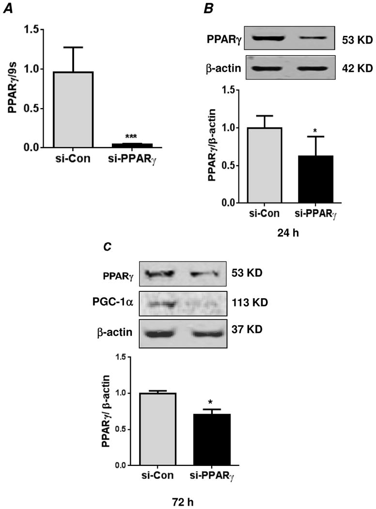
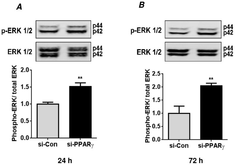
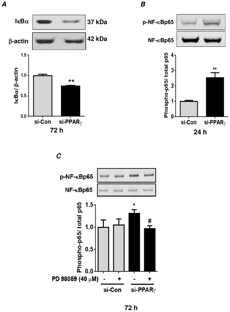
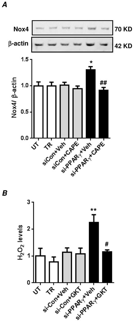
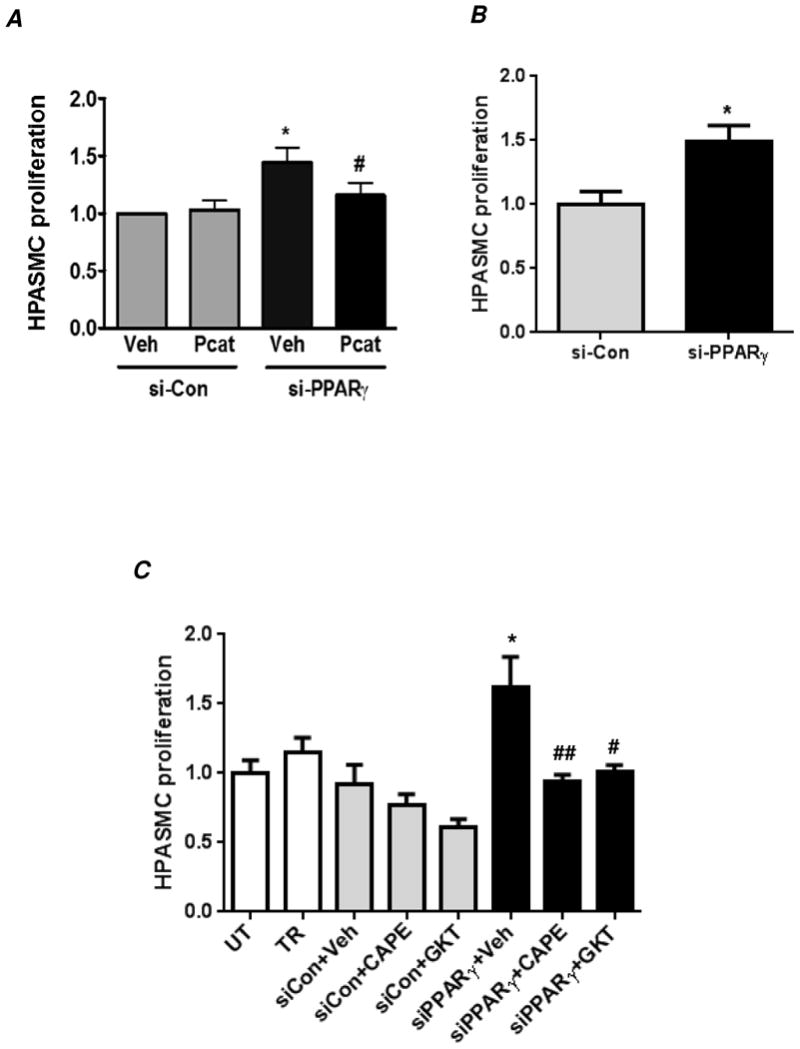
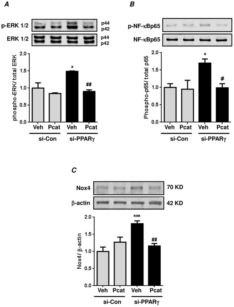
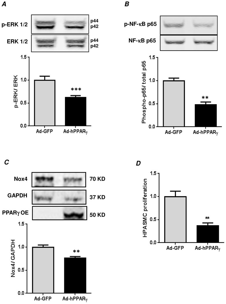
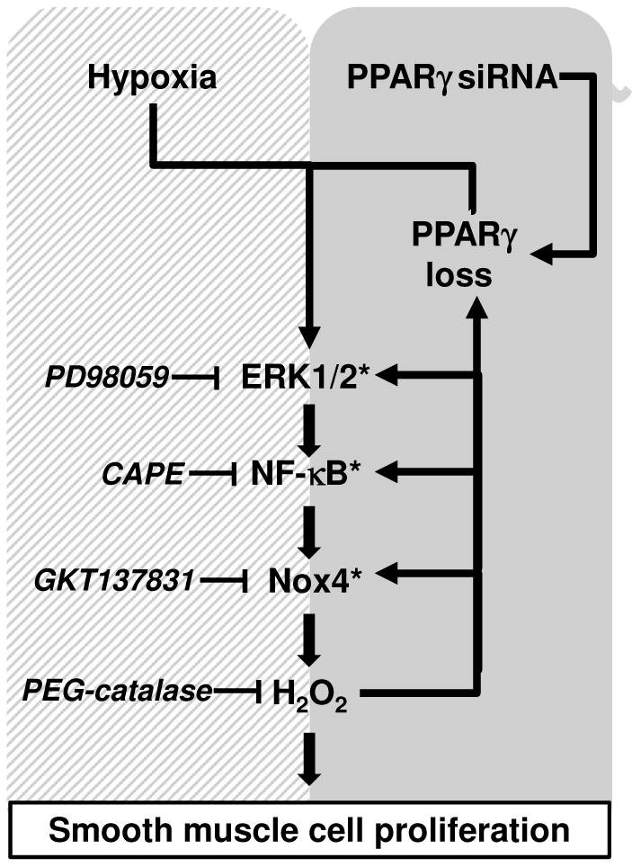
Hypoxia stimulates pulmonary hypertension (PH) in part by increasing the proliferation of pulmonary vascular wall cells. Recent evidence suggests that signaling events involved in hypoxia-induced cell proliferation include sustained nuclear factor-kappaB (NF-κB) activation, increased NADPH oxidase 4 (Nox4) expression, and downregulation of peroxisome proliferator-activated receptor gamma (PPARγ) levels. To further understand the role of reduced PPARγ levels associated with PH pathobiology, siRNA was employed to reduce PPARγ levels in human pulmonary artery smooth muscle cells (HPASMC) in vitro under normoxic conditions. PPARγ protein levels were reduced to levels comparable to those observed under hypoxic conditions. Depletion of PPARγ for 24-72 h activated mitogen-activated protein kinase, ERK 1/2, and NF-κB. Inhibition of ERK 1/2 prevented NF-κB activation caused by PPARγ depletion, indicating that ERK 1/2 lies upstream of NF-κB activation. Depletion of PPARγ for 72 h increased NF-κB-dependent Nox4 expression and H2O2 production. Inhibition of NF-κB or Nox4 attenuated PPARγ depletion-induced HPASMC proliferation. Degradation of PPARγ depletion-induced H2O2 by PEG-catalase prevented HPASMC proliferation and also ERK 1/2 and NF-κB activation and Nox4 expression, indicating that H2O2 participates in feed-forward activation of the above signaling events. Contrary to the effects of PPARγ depletion, HPASMC PPARγ overexpression reduced ERK 1/2 and NF-κB activation, Nox4 expression, and cell proliferation. Taken together these findings provide novel evidence that PPARγ plays a central role in the regulation of the ERK1/2-NF-κB-Nox4-H2O2 signaling axis in HPASMC. These results indicate that reductions in PPARγ caused by pathophysiological stimuli such as prolonged hypoxia exposure are sufficient to promote the proliferation of pulmonary vascular smooth muscle cells observed in PH pathobiology.

Keywords: ERK 1/2; NF-κB; Nox4; PPARγ; Pulmonary artery smooth muscle cell; Pulmonary hypertension.


Figures

Figure 1
Figure 1. siRNA-mediated knockdown of PPARγ
(A) Human pulmonary artery smooth muscle cells (HPASMC) were transfected with control siRNA (si-Con) or PPARγ siRNA (si-PPARγ) under normoxic conditions (21% O2 and 5% CO2 at 37°C) for 72 hours. RNA was isolated, and PPARγ mRNA levels were determined by qRT-PCR. The mRNA levels were normalized to 9S ribosomal RNA. Each bar represents the mean ± SEM of levels of PPARγ mRNA relative to 9S ribosomal RNA in the same sample expressed as fold-change versus control. n=6, ***p<0.001. In related experiments, HPASMC were transfected with control siRNA or PPARγ siRNA under normoxic conditions for 24 hours (B) or 72 hours (C). Cells were lysed and analyzed for PPARγ protein levels or its target gene, PGC-1α by western blot. The PPARγ protein levels were normalized with β-actin levels. Each bar represents the mean ± SEM of levels of PPARγ protein relative to β-actin in the same sample expressed as fold-change versus control. n=3, *p<0.05.
Figure 2
Figure 2. PPARγ knockdown promotes HPASMC ERK 1/2 activation
HPASMC were transfected with control siRNA (si-Con) or PPARγ siRNA (si-PPARγ) under normoxic conditions for 24 hours (A) or 72 hours (B). Cells were lysed and analyzed by western blot for activation of extracellular regulated kinase (ERK)-1/2 using an anti-phospho-(Thr202/Tyr204)-ERK1/2 antibody. The phospho-ERK 1/2 levels were normalized with total ERK 1/2 levels. Each bar represents levels of phospho-ERK 1/2 relative to total ERK 1/2 in the same sample expressed as fold-change versus control. n=3, **p<0.01.
Figure 3
Figure 3. PPARγ knockdown in HPASMC promotes ERK 1/2-mediated activation of NF-κB
HPASMC were transfected with control siRNA (si-Con) or PPARγ siRNA (si-PPARγ) under normoxic conditions for 72 hours (A) or 24 hours (B). Cells were lysed and analyzed by western blot for IκBα (A) or phospho-p65 using an anti-phospho-(Ser536)-NF-κBp65 antibody (B). In (A), the levels of IκBα were normalized with β-actin. Each bar represents mean ± SEM IKBα levels relative to β-actin in the same sample expressed as fold-change versus control. n=3, **p<0.01. In (B), phospho-NF-κBp65 levels were normalized with total NF-κBp65 levels. Each bar represents mean ± SEM of phospho-NF-κBp65 levels relative to total NF-κBp65 in the same sample expressed as fold-change versus control. n=3-6, **p<0.01. (C) Cells were transfected with siRNA as described above, and treated with the ERK 1/2 inhibitor, PD98059 (40 μM) or vehicle (DMSO) during the final 24 hours of the 72 hour transfection period. Cells were lysed and analyzed for phospho-NF-κBp65 by western blot as described above. Each bar represents mean ± SEM phospho-NF-κBp65 levels relative to total NF-κBp65 in the same sample expressed as fold-change versus control. n=3, *p<0.05 vs si-Con; #p<0.05 vs vehicle treated si-PPARγ.
Figure 4
Figure 4. PPARγ knockdown promotes NF-κB-dependent Nox4 expression and Nox4-derived H2O2 generation in HPASMC
(A) HPASMC were untransfected (UT) or transfected with transfection reagent alone (TR), control siRNA (si-Con) or PPARγ siRNA (si-PPARγ) under normoxic conditions for 72 hours. Cells were treated with CAPE compound (10 μM) or equivalent volume of DMSO (vehicle) during the final 24 hours of the 72 hours transfection period. Cells were lysed and analyzed by western blot for Nox4 protein expression. Nox4 levels were normalized with β-actin levels. Each bar represents mean ± SEM Nox4 relative to β-actin in the same sample expressed as fold-change versus UT. n=6-7, *p<0.05 compared to UT, ##p<0.01 compared to PPARγ siRNA transfected cells that were treated with DMSO. (B) HPASMC were transfected as above under normoxic conditions for 72 hours. Cells were treated with GKT137831 (20 μM) or equivalent volume of DMSO (vehicle) during the final 24 hours of the 72 hour transfection period, and H2O2 concentration was measured with the Amplex Red assay. Each bar represents mean ± SEM H2O2 concentration as fold-change versus UT. n=6-7, **p<0.01 compared to UT; #p<0.05 compared to si-PPARγ+Veh.
Figure 5
Figure 5. PPARγ knockdown promotes HPASMC proliferation via an NF-κB-Nox4-H2O2-dependent mechanism
HPASMC were untransfected (UT) or transfected with transfection reagent alone (TR), control siRNA (si-Con) or PPARγ siRNA (si-PPARγ) under normoxic conditions for 72 hours. Selected HPASMC were treated with PEG-catalase (1000 U/ml) (A), CAPE (10 μM) or GKT137831 (20 μM) (C) or with vehicle (Veh, DMSO) during the last 24 hours of the 72 hour transfection period. Cell proliferation was determined by MTT assay. (B) Cell proliferation upon PPARγ depletion was also verified by manual cell counting method. In (A), each bar represents mean ± SEM HPASMC proliferation as fold-change versus control. n=3, *p<0.05 compared to Veh/si-Con; #p<0.05 compared to Veh/si-PPARγ. In (B), each bar represents mean ± SEM HPASMC proliferation (cells/ml) as fold-change versus control. n=3, *p<0.05 compared to si-Con. In (C), each bar represents mean ± SEM HPASMC proliferation as fold-change versus UT. n=5-7, **p<0.01 compared to UT; # ##p<0.05 and 0.01, respectively, compared to si-PPARγ+Veh.
Figure 6
Figure 6. Production of H2O2 caused by PPARγ knockdown is required for sustained ERK 1/2 and NF-κB activation as well as Nox4 expression
HPASMC were transfected with control siRNA (si-Con) or PPARγ siRNA (si-PPARγ) under normoxic conditions for 72 hours. During final 24 hours of transfection, cells were treated with PEG-catalase (Pcat, 1000 U/ml) or with DMSO as control vehicle (Veh). Cell lysates were immunoblotted for phospho-ERK 1/2 (A) phospho-p65 (B) or Nox4 (C). In (A), each bar represents mean ± SEM phospho-ERK 1/2 relative to total ERK 1/2 in the same sample expressed as fold-change versus control. n=3, *p<0.05 vs si-Con; ##p<0.01 vs vehicle treated si-PPARγ. In (B), each bar represents mean ± SEM phospho-NF-κBp65 relative to total NF-κBp65 in the same sample expressed as fold-change versus control. n=3, *p<0.05 vs si-Con; #p<0.05 vs vehicle treated si-PPARγ. In (C), each bar represents mean ± SEM Nox4 relative to β-actin in the same sample expressed as fold-change versus control. n=6, ***p<0.001 vs si-Con; ##p<0.01 vs vehicle treated si-PPARγ.
Figure 7
Figure 7. PPARγ overexpression reduces basal ERK 1/2 and NF-κB activation and Nox4 protein expression and HPASMC proliferation
Confluent HPASMC monolayers were transfected with human PPARγ in adenovirus (Ad-hPPARγ) or Ad-GFP (Vector Biolabs, Philadelphia, PA) as described in Materials and Methods. Cells were lysed and immunoblotted for phospho-ERK 1/2 (A), phospho-p65 (B), or Nox4 (C). In (D), HPASMC proliferation was measured by manual cell counting method. In (A), each bar represents mean ± SEM phospho-ERK 1/2 relative to total ERK 1/2 in the same sample expressed as fold-change versus Ad-GFP. n=4, ***p<0.001. In (B), each bar represents mean ± SEM phospho-p65 relative to total p65 in the same sample expressed as fold-change versus Ad-GFP. n=4, **p<0.01. In (C), each bar represents mean ± SEM Nox4 relative to GAPDH in the same sample expressed as fold-change versus Ad-GFP. n=4, **p<0.01. Overexpression of PPARγ (PPARγ-OE) was confirmed by immunoblotting. The image was captured at a lower intensity to highlight the expression of exogenously introduced PPARγ. In (D), each bar represents mean ± SEM HPASMC proliferation (cells/ml) expressed as fold-change versus Ad-GFP. n=4, **p<0.01. Similar results were obtained by MTT assay (data not shown).
Figure 8
Figure 8. Schematic representation of ERK1/2-p65-Nox4-mediated pathways of cell proliferation in HPASMC caused by PPARγ depletion
We previously demonstrated that hypoxia promotes HPASMC proliferation and PPARγ downregulation via an ERK1/2-NF-κB p65-Nox4-dependent mechanism [19] (hatched box). The findings in the current study (gray box) demonstrate that loss of PPARγ is sufficient to activate ERK1/2 and increase NF-κB p65 activity, Nox4 expression, H2O2 production, and HPASMC proliferation whereas PPARγ overexpression attenuated ERK1/2, NF-κB, and Nox4 (denoted by asterisks). These findings indicate that PPARγ provides constitutive inhibition of ERK, NF-κB, and Nox4 signaling pathways critical in PASMC proliferation. These findings further emphasize that targeting PPARγ may provide a useful therapeutic strategy to attenuate PASMC proliferation and pulmonary vascular remodeling.

References

    1. Picard F, Auwerx J. PPAR(gamma) and glucose homeostasis. Annu Rev Nutr. 2002;22:167–197. - PubMed
    1. Ahmadian M, Suh JM, Hah N, Liddle C, Atkins AR, Downes M, Evans RM. PPARγ signaling and metabolism: the good, the bad and the future. Nat Med. 2013;19:557–566. - PMC - PubMed
    1. Bardot O, Aldridge TC, Latruffe N, Green S. PPAR-RXR heterodimer activates a peroxisome proliferator response element upstream of the bifunctional enzyme gene. Biochem Biophys Res Commun. 1993;192:37–45. - PubMed
    1. Tontonoz P, Hu E, Spiegelman BM. Stimulation of adipogenesis in fibroblasts by PPAR gamma 2, a lipid-activated transcription factor. Cell. 1994;79:1147–1156. - PubMed
    1. Pascual G, Fong AL, Ogawa S, Gamliel A, Li AC, Perissi V, Rose DW, Willson TM, Rosenfeld MG, Glass CK. A SUMOylation-dependent pathway mediates transrepression of inflammatory response genes by PPAR-gamma. Nature. 2005;437:759–763. - PMC - PubMed

Publication types

MeSH terms

LinkOut - more resources