

Decompressive craniectomy reduces white matter injury after controlled cortical impact in mice
- PMID: 25557588
- PMCID: PMC4449625
- DOI: 10.1089/neu.2014.3564
Decompressive craniectomy reduces white matter injury after controlled cortical impact in mice
Abstract
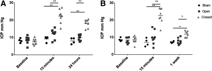
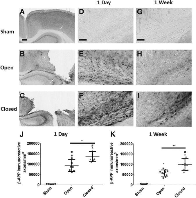
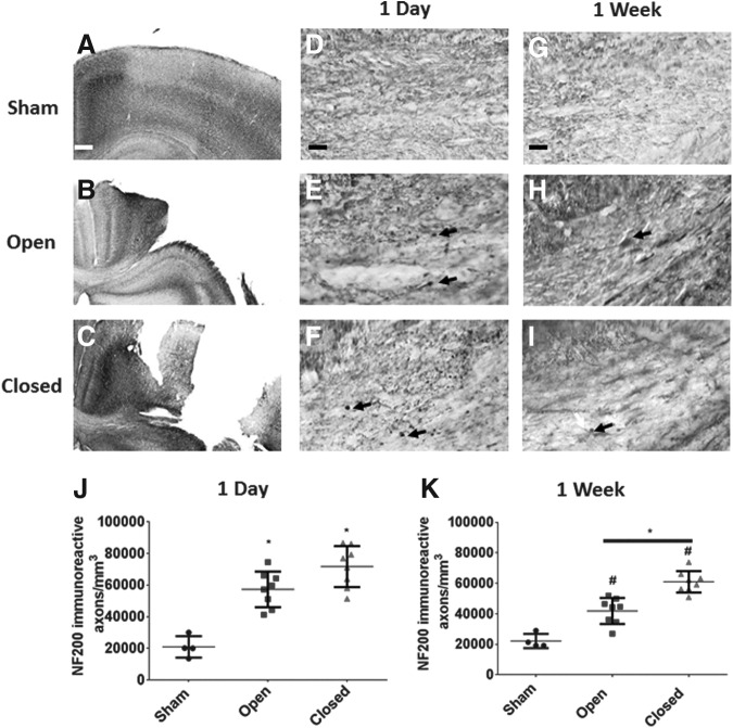
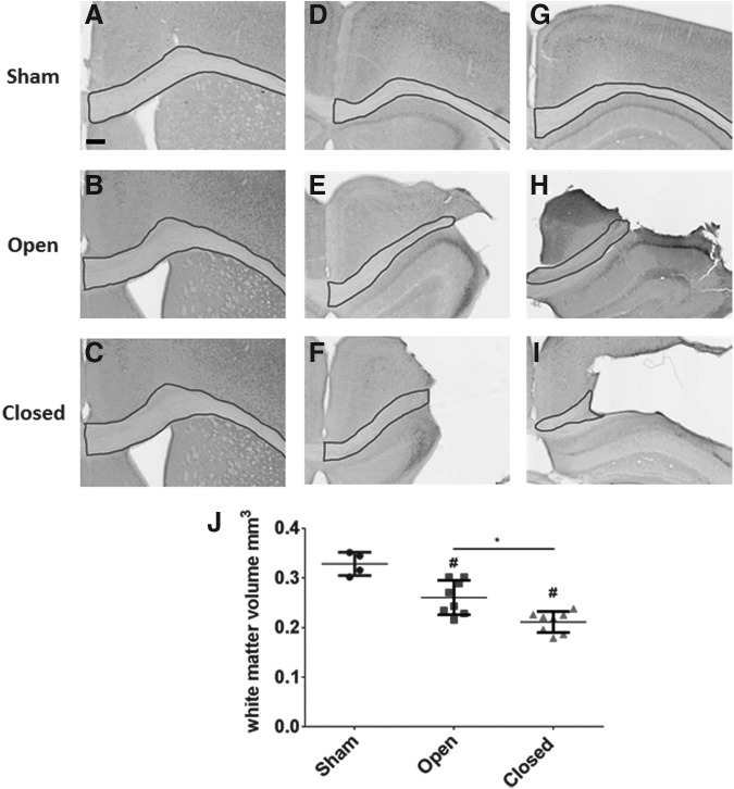
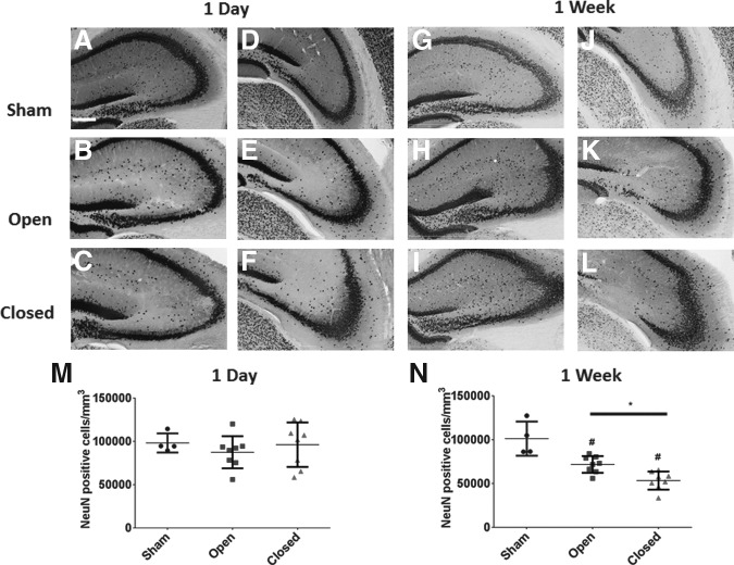
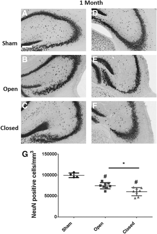
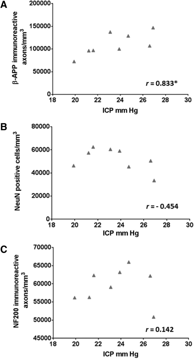
Reduction and avoidance of increases in intracranial pressure (ICP) after severe traumatic brain injury (TBI) continue to be the mainstays of treatment. Traumatic axonal injury is a major contributor to morbidity after TBI, but it remains unclear whether elevations in ICP influence axonal injury. Here we tested the hypothesis that reduction in elevations in ICP after experimental TBI would result in decreased axonal injury and white matter atrophy in mice. Six-week-old male mice (C57BL/6J) underwent either moderate controlled cortical impact (CCI) (n=48) or Sham surgery (Sham, n=12). Immediately after CCI, injured animals were randomized to a loose fitting plastic cap (Open) or replacement of the previously removed bone flap (Closed). Elevated ICP was observed in Closed animals compared with Open and Sham at 15 min (21.4±4.2 vs. 12.3±2.9 and 8.8±1.8 mm Hg, p<0.0001) and 1 day (17.8±3.7 vs. 10.6±2.0 and 8.9±1.9 mm Hg, p<0.0001) after injury. Beta amyloid precursor protein staining in the corpus callosum and ipsilateral external capsule revealed reduced axonal swellings and bulbs in Open compared with Closed animals (32% decrease, p<0.01 and 40% decrease, p<0.001 at 1 and 7 days post-injury, respectively). Open animals were also found to have decreased neurofilament-200 stained axonal swellings at 7 days post-injury compared with Open animals (32% decrease, p<0.001). At 4 weeks post-injury, Open animals had an 18% reduction in white matter volume compared with 34% in Closed animals (p<0.01). Thus, our results indicate that CCI with decompressive craniectomy was associated with reductions in ICP and reduced pericontusional axonal injury and white matter atrophy. If similar in humans, therapeutic interventions that ameliorate intracranial hypertension may positively influence white matter injury severity.
Keywords: axonal injury; controlled cortical impact; intracranial pressure; traumatic brain injury; white matter.
Figures
Similar articles
-
Effect of early and delayed decompressive craniectomy on secondary brain damage after controlled cortical impact in mice.J Neurotrauma. 2006 Jul;23(7):1083-93. doi: 10.1089/neu.2006.23.1083. J Neurotrauma. 2006. PMID: 16866621
-
Bilateral decompressive craniectomy for patients with malignant diffuse brain swelling after severe traumatic brain injury: a 37-case study.J Neurotrauma. 2010 Feb;27(2):341-7. doi: 10.1089/neu.2009.1040. J Neurotrauma. 2010. PMID: 19715392
-
Quantitative structural changes in white and gray matter 1 year following traumatic brain injury in rats.Acta Neuropathol. 2002 Jun;103(6):607-14. doi: 10.1007/s00401-001-0510-8. Epub 2002 Mar 20. Acta Neuropathol. 2002. PMID: 12012093
-
Decompressive craniectomy: a meta-analysis of influences on intracranial pressure and cerebral perfusion pressure in the treatment of traumatic brain injury.J Neurosurg. 2012 Sep;117(3):589-96. doi: 10.3171/2012.6.JNS101400. Epub 2012 Jul 13. J Neurosurg. 2012. PMID: 22794321 Review.
-
Decompressive craniectomy in traumatic brain injury after the DECRA trial. Where do we stand?Curr Opin Crit Care. 2013 Apr;19(2):101-6. doi: 10.1097/MCC.0b013e32835eba1a. Curr Opin Crit Care. 2013. PMID: 23422159 Review.
Cited by
-
A Systematic Review of Traumatic Brain Injury in Modern Rodent Models: Current Status and Future Prospects.Biology (Basel). 2024 Oct 11;13(10):813. doi: 10.3390/biology13100813. Biology (Basel). 2024. PMID: 39452122 Free PMC article. Review.
-
Prolonged course of brain edema and neurological recovery in a translational model of decompressive craniectomy after closed head injury in mice.Front Neurol. 2023 Nov 20;14:1308683. doi: 10.3389/fneur.2023.1308683. eCollection 2023. Front Neurol. 2023. PMID: 38053795 Free PMC article.
-
Animal models of traumatic brain injury: a review of pathophysiology to biomarkers and treatments.Exp Brain Res. 2021 Oct;239(10):2939-2950. doi: 10.1007/s00221-021-06178-6. Epub 2021 Jul 29. Exp Brain Res. 2021. PMID: 34324019 Review.
-
Delayed Hypoxemia after Traumatic Brain Injury Exacerbates Long-Term Behavioral Deficits.J Neurotrauma. 2018 Mar 1;35(5):790-801. doi: 10.1089/neu.2017.5354. Epub 2018 Jan 12. J Neurotrauma. 2018. PMID: 29149808 Free PMC article.
-
White Matter Injury in Early Brain Injury after Subarachnoid Hemorrhage.Cell Transplant. 2019 Jan;28(1):26-35. doi: 10.1177/0963689718812054. Epub 2018 Nov 16. Cell Transplant. 2019. PMID: 30442028 Free PMC article. Review.
References
-
- Graham D.I., Adams J.H., Murray L.S., and Jennett B. (2005). Neuropathology of the vegetative state after head injury. Neuropsychol. Rehabil. 15, 198–213 - PubMed
-
- Gennarelli T.A., Thibault L.E., Adams J.H., Graham D.I., Thompson C.J., and Marcincin R.P. (1982). Diffuse axonal injury and traumatic coma in the primate. Ann. Neurol. 12, 564–574 - PubMed
-
- Smith D.H., Meaney D.F., and Shull W.H. (2003). Diffuse axonal injury in head trauma. J. Head Trauma Rehabil. 18, 307–316 - PubMed
-
- Smith D.H., Chen X.H., Iwata A., and Graham D.I. (2003). Amyloid beta accumulation in axons after traumatic brain injury in humans. J. Neurosurg. 98, 1072–1077 - PubMed
-
- Povlishock J.T., Erb D.E., and Astruc J. (1992). Axonal response to traumatic brain injury: reactive axonal change, deafferentation, and neuroplasticity. J. Neurotrauma 9, Suppl 1, S189–S200 - PubMed
Publication types
MeSH terms
Grants and funding
LinkOut - more resources
Full Text Sources
Other Literature Sources
Miscellaneous
