. 2015 Jan 8;96(1):153-61.
doi: 10.1016/j.ajhg.2014.11.014. Epub 2014 Dec 31.

Defects of CRB2 cause steroid-resistant nephrotic syndrome

Affiliations

Defects of CRB2 cause steroid-resistant nephrotic syndrome

Lwaki Ebarasi et al. Am J Hum Genet. .

Abstract

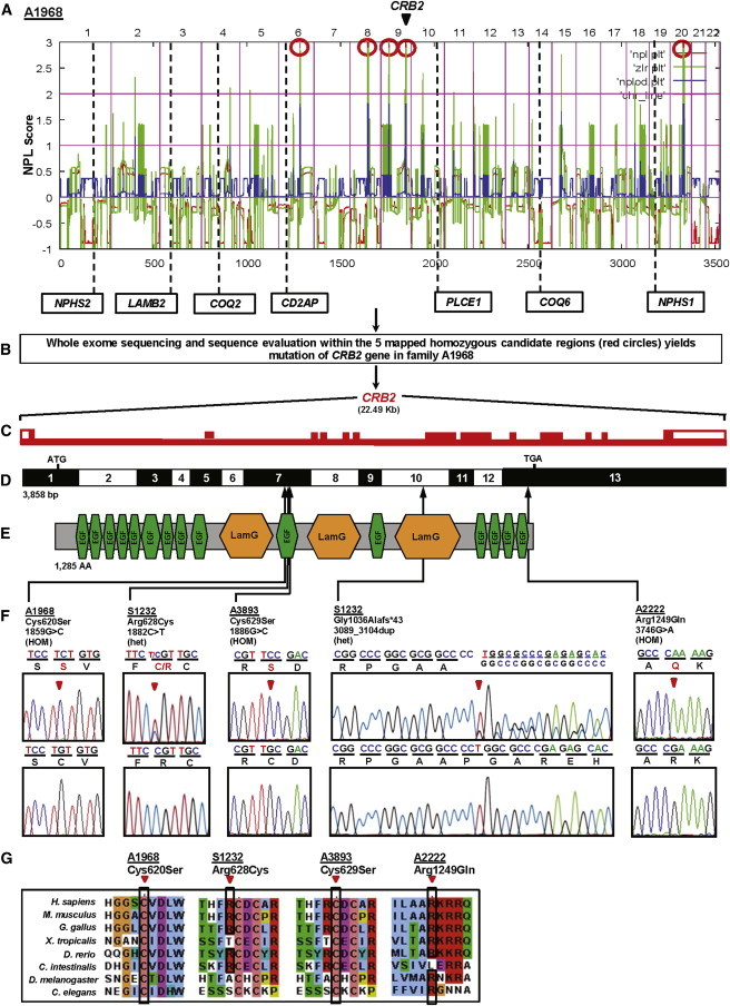
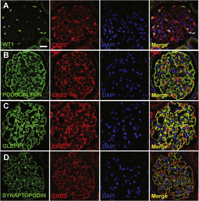
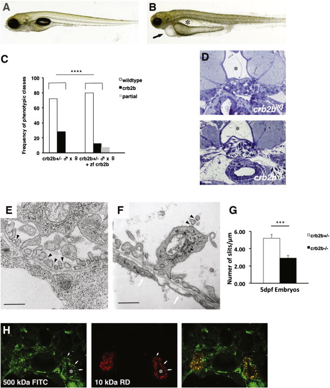
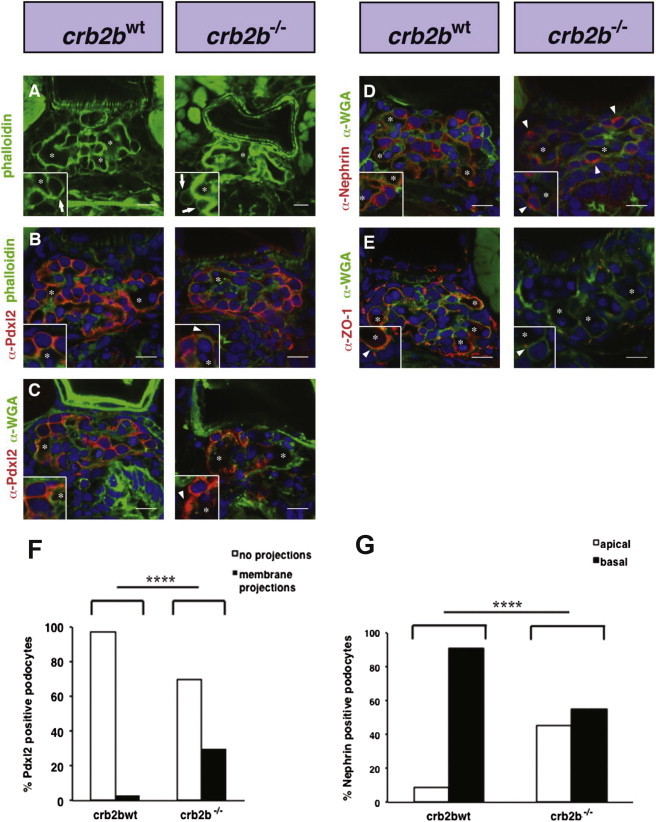
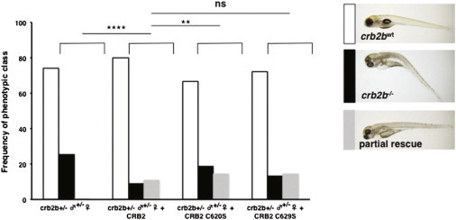
Nephrotic syndrome (NS), the association of gross proteinuria, hypoalbuminaemia, edema, and hyperlipidemia, can be clinically divided into steroid-sensitive (SSNS) and steroid-resistant (SRNS) forms. SRNS regularly progresses to end-stage renal failure. By homozygosity mapping and whole exome sequencing, we here identify recessive mutations in Crumbs homolog 2 (CRB2) in four different families affected by SRNS. Previously, we established a requirement for zebrafish crb2b, a conserved regulator of epithelial polarity, in podocyte morphogenesis. By characterization of a loss-of-function mutation in zebrafish crb2b, we now show that zebrafish crb2b is required for podocyte foot process arborization, slit diaphragm formation, and proper nephrin trafficking. Furthermore, by complementation experiments in zebrafish, we demonstrate that CRB2 mutations result in loss of function and therefore constitute causative mutations leading to NS in humans. These results implicate defects in podocyte apico-basal polarity in the pathogenesis of NS.


Figures

Figure 1
Figure 1
Homozygosity Mapping and WES Identifies CRB2 Mutations as Causing Steroid-Resistant Nephrotic Syndrome in Humans (A) Nonparametric LOD score (NPL) profile across the human genome in two sibs with SRNS of consanguineous family A1968. SNP mapping was performed with the Affymetrix 250 StyI array. SNP positions on human chromosomes are concatenated from p-ter (left) to q-ter (right) on the x axis. Genetic distance is given in cM. Five maximum NPL peaks (red circles) indicate candidate regions of homozygosity by descent. Note that none of the peaks overlap with any of the seven known recessive NS loci. (B) WES of one of the affected siblings from family A1968 and sequence evaluation within the five mapped homozygous candidate regions (red circles in A) yields mutation of CRB2 in A1968. (C) The CRB2 gene extends over 22.49 kb and contains 13 exons (vertical hatches). (D) Exon structure of human CRB2 cDNA. Positions of start codon (ATG) at nt +1 and of stop codon (TGA) are indicated. For the mutations detected (see F), arrows indicate positions in relation to exons and protein domains (see E). (E) Domain structure of the CRB2 protein. 15 EGF-like; calcium-binding domains (green) and 3 Laminin G-like domains (orange) are predicted. (F) CRB2 mutations detected in four families affected by SRNS. Family number and predicted translational change are indicated (see Table 1). Sequence traces are shown for homozygous mutations above normal controls, and mutated nucleotides are indicated by arrowheads. “HOM” denotes homozygous and “het” denotes heterozygous mutations. (G) The conservation across evolution of altered amino acid residues is shown for all four missense variants (p.Cys620Ser, p.Arg628Cys, p.Cys629Ser, and p.Arg1249Gln).
Figure 2
Figure 2
Localization of CRB2 in Adult Rat Kidney (A) Coimmunofluorescence of CRB2 (Abgent) with WT1 (Santa Cruz Biotech). CRB2 localizes to podocytes, the nuclei of which are marked by WT1. (B–D) Coimmunofluorescence of CRB2 with podocytic markers PODOCALYXIN (B), GLEPP1 (C), and SYNAPTOPODIN (D) (American Research Products). CRB2 colocalizes most tightly with GLEPP1 among podocytic markers used in immunofluorescence. Note that PODOCALYXIN and GLEPP1 mark the apical podocyte foot process domain, and GLEPP1 is next to the slit membrane adherens junctions. SYNAPTOPODIN marks podocyte processes distal of the slit membrane. Scale bar represents 10 μm. PODOCALYXIN and GLEPP1 antibodies were kindly provided by Roger C. Wiggins at the University of Michigan.
Figure 3
Figure 3
Zebrafish crb2b−/− Mutants Have Morphologically Defective Podocytes (A and B) Brightfield images of zebrafish 5 days postfertilization (dpf): crb2bwt (A) and crb2b−/− larvae (B). crb2b−/− homozygous mutants show reduced eye size, pericardial effusion (arrow), and pronephric cysts (asterisk). (C) Rescue of the crb2b−/− eye, pronephric, and pericardial effusion phenotypes by injection of full-length zebrafish Crb2b mRNA. (crb2b+/− ♂ × ♀, n = 75; crb2b+/− ♂ × ♀ + zebrafish Crb2b, n = 183 embryos, p < 0.0001). Complete rescue (black bars) and partial rescue (gray bars) frequencies are shown. Phenotypes were scored at 4.5 dpf. (D) Transverse sections at the level of the glomerulus in crb2bwt and crb2b−/− 5 dpf larvae. In controls, the glomerulus is directly ventral to the notochord (asterisk) and dorsal aorta. In crb2bwt, capillary loops are densely packed and covered with podocytes. In crb2b−/− glomeruli, capillary loops are fused together and podocytes are attached to the loops. (E and F) Electron microsopic analysis of podocyte foot process organization in control crb2bwt (E) and crb2b−/− (F) homozygotes at 5 dpf. In crb2bwt, slit diaphragms are visible (black arrowheads in E). The crb2b−/− mutant podocytes show disorganized foot process formation, apical membrane projections containing slit diaphragms (black arrowheads in F) in the urinary space, and a rarefaction of slit diaphragms. A glomerular basement membrane is visible. The endothelium lacks membrane fenestrations in crb2b−/− mutants (white arrowheads in F). Scale bars represent 500 nm. (G) Quantification of slit diaphragm defects in crb2b−/− mutants (p < 0.001, n = 3 regions/glomerulus from 3 different glomeruli). Data represent the mean ± SEM. (H) Dye filtration assay shows that FITC-labeled 500 kDa (green) and rhodamine-labeled 10 kDa dextran (red) dyes injected into living 4.5 dpf crb2b−/− mutants are both passed into and endocytosed (arrows) by the pronephric proximal tubules. The asterisk marks the tubule lumen.
Figure 4
Figure 4
Apical Basal Polarity Is Affected in Zebrafish crb2b−/− Glomerular Podocytes (A) Phalloidin staining outlines the dense podocyte actin foot process network surrounding capillary lumens. In crb2b−/− mutants, capillary lumens are not compartmentalized but fused into larger vessels. Insets show enlarged images of podocytes. Asterisks mark glomerular capillary lumens. (B) In crb2bwt, α-Pdxl2 localizes to apical podocyte membranes. In crb2b−/− mutant podocytes, α-Pdxl2 staining is found in ectopic apical projections (arrowheads). (C) Glomerular basement membranes are visualized by α-wheat germ agglutinin (α-WGA) staining. Asterisks mark glomerular capillary lumens. Pdxl2 staining is again found in ectopic apical projections in crb2b−/− mutant podocytes (arrowheads). (D) α-WGA shows glomerular basement membranes. α-Nephrin staining is basally localized in control podocytes but apically mislocalized in crb2b−/− podocytes (arrowheads). (E) α-ZO-1 (Zymed) podocyte staining lines the GBM in crb2bwt but is diminished in crb2b−/− mutants. Scale bars represent 10 μm. (F) Quantification of α-Pdxl2-positive membrane projections in crb2bwt and crb2b−/− podocytes. n = 37 (tallied from 6 embryos) crb2bwt control and n = 30 (tallied from 4 embryos) crb2b−/− podocytes. (G) Quantification of α-Nephrin localization in crb2bwt and crb2b−/− podocytes. n = 106 (tallied from 9 embryos) control and n = 67 (tallied from 5 embryos) crb2b−/− podocytes. p < 0.0001.
Figure 5
Figure 5
Functional Assay of Human CRB2 Mutations in Zebrafish crb2b+/− ♂ × ♀ Incrosses Phenotypic frequencies of crb2bwt and crb2b−/− mutant embryos after injection of human CRB2 control mRNA and the mRNAs harboring the mutations c.1859G>C; CRB2C620S and c.1882C>T; CRB2C629S (crb2b+/− ♂ × ♀, n = 363; crb2b+/− ♂ × ♀ + CRB2, n = 167, p = 0.02; crb2b+/− ♂ × ♀ + CRB2C620S, n = 117, p < 0.0001; crb2b+/− ♂ × ♀ + CRB2C629S, n = 283, p = 0.06; ns, no significant difference). Phenotypic classes of embryos recovered from rescue experiments. Partially rescued embryos have a straight body axis, phenotypically wild-type eyes, and lack pronephric cysts but still show some pericardial effusion. No rescue (black bars) and partial rescue (gray bars) frequencies are shown. Phenotypes were scored at 4.5 dpf.

References

    1. Reiser J., Kriz W., Kretzler M., Mundel P. The glomerular slit diaphragm is a modified adherens junction. J. Am. Soc. Nephrol. 2000;11:1–8. - PubMed
    1. Wiggins R.C. The spectrum of podocytopathies: a unifying view of glomerular diseases. Kidney Int. 2007;71:1205–1214. - PubMed
    1. Mekahli D., Liutkus A., Ranchin B., Yu A., Bessenay L., Girardin E., Van Damme-Lombaerts R., Palcoux J.B., Cachat F., Lavocat M.P. Long-term outcome of idiopathic steroid-resistant nephrotic syndrome: a multicenter study. Pediatr. Nephrol. 2009;24:1525–1532. - PubMed
    1. ISKDC Primary nephrotic syndrome in children: clinical significance of histopathologic variants of minimal change and of diffuse mesangial hypercellularity. A Report of the International Study of Kidney Disease in Children. Kidney Int. 1981;20:765–771. - PubMed
    1. Benoit G., Machuca E., Antignac C. Hereditary nephrotic syndrome: a systematic approach for genetic testing and a review of associated podocyte gene mutations. Pediatr. Nephrol. 2010;25:1621–1632. - PMC - PubMed

Publication types

MeSH terms