

Syringaldehyde exerts neuroprotective effect on cerebral ischemia injury in rats through anti-oxidative and anti-apoptotic properties
- PMID: 25558237
- PMCID: PMC4281426
- DOI: 10.4103/1673-5374.145353
Syringaldehyde exerts neuroprotective effect on cerebral ischemia injury in rats through anti-oxidative and anti-apoptotic properties
Abstract
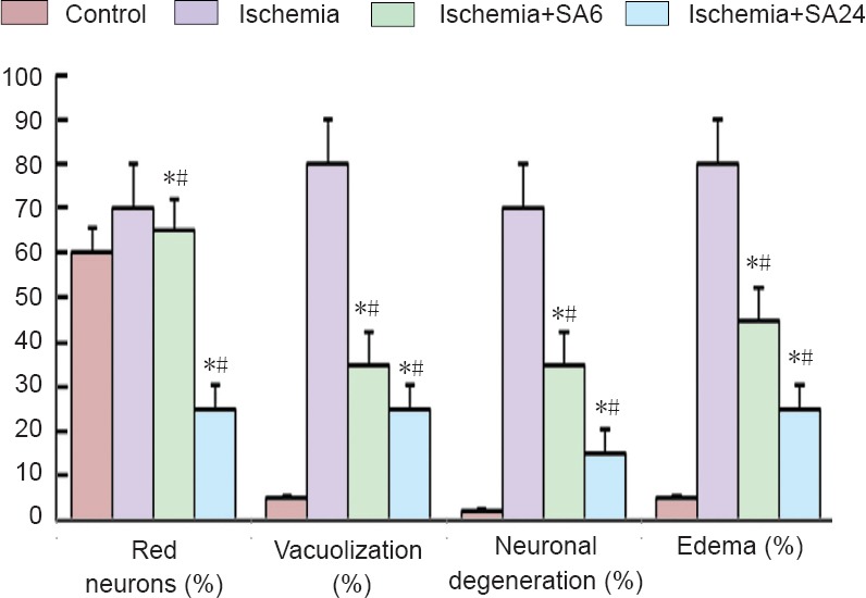
There are few studies on the neuroprotective effects of syringaldehyde in a rat model of cerebral ischemia. The study aimed to elucidate the mechanisms underlying the neuroprotective effects of syringaldehyde on ischemic brain cells. Rat models of cerebral ischemia were intraperitoneally administered syringaldehyde. At 6 and 24 hours after syringaldehyde administration, cell damage in the brain of cerebral ischemia rats was obviously reduced, superoxide dismutase activity and nuclear respiratory factor 1 expression in the brain tissue were markedly increased, malondiadehyde level was obviously decreased, apoptosis-related cysteine peptidase caspase-3 and -9 immunoreactivity was obviously decreased, and neurological function was markedly improved. These findings suggest that syringaldehyde exerts neuroprotective effects on cerebral ischemia injury through anti-oxidation and anti-apoptosis.
Keywords: apoptosis; brain ischemia; inflammatory; nerve regeneration; neural regeneration; neuroprotective effects; oxidative stress; syringaldehyde.
Conflict of interest statement
Figures


Similar articles
-
Neuroprotective effects of daidzein on focal cerebral ischemia injury in rats.Neural Regen Res. 2015 Jan;10(1):146-52. doi: 10.4103/1673-5374.150724. Neural Regen Res. 2015. PMID: 25788936 Free PMC article.
-
SC79, the AKT Activator Protects Cerebral Ischemia in a Rat Model of Ischemia/Reperfusion Injury.Med Sci Monit. 2018 Aug 3;24:5391-5397. doi: 10.12659/MSM.910191. Med Sci Monit. 2018. PMID: 30074018 Free PMC article.
-
Neuroprotective effects of salidroside on focal cerebral ischemia/reperfusion injury involve the nuclear erythroid 2-related factor 2 pathway.Neural Regen Res. 2015 Dec;10(12):1989-96. doi: 10.4103/1673-5374.172317. Neural Regen Res. 2015. PMID: 26889188 Free PMC article.
-
Neuroprotective and anti-apoptotic effects of liraglutide in the rat brain following focal cerebral ischemia.Neuroscience. 2014 Dec 5;281:269-81. doi: 10.1016/j.neuroscience.2014.09.064. Epub 2014 Oct 6. Neuroscience. 2014. PMID: 25301749
-
Senkyunolide I protects rat brain against focal cerebral ischemia-reperfusion injury by up-regulating p-Erk1/2, Nrf2/HO-1 and inhibiting caspase 3.Brain Res. 2015 Apr 24;1605:39-48. doi: 10.1016/j.brainres.2015.02.015. Epub 2015 Feb 16. Brain Res. 2015. PMID: 25698615
Cited by
-
Acetylcholinesterase Inhibitory and Antioxidant Activity of the Compounds Isolated from Vanda roxburghii.Adv Pharmacol Pharm Sci. 2021 Mar 27;2021:5569054. doi: 10.1155/2021/5569054. eCollection 2021. Adv Pharmacol Pharm Sci. 2021. PMID: 33855299 Free PMC article.
-
Merging the Multi-Target Effects of Phytochemicals in Neurodegeneration: From Oxidative Stress to Protein Aggregation and Inflammation.Antioxidants (Basel). 2020 Oct 20;9(10):1022. doi: 10.3390/antiox9101022. Antioxidants (Basel). 2020. PMID: 33092300 Free PMC article. Review.
-
Study on the Neuroprotective, Radical-Scavenging and MAO-B Inhibiting Properties of New Benzimidazole Arylhydrazones as Potential Multi-Target Drugs for the Treatment of Parkinson's Disease.Antioxidants (Basel). 2022 Apr 29;11(5):884. doi: 10.3390/antiox11050884. Antioxidants (Basel). 2022. PMID: 35624746 Free PMC article.
-
Syringaldehyde Exhibits Antibacterial and Antioxidant Activities against Mycobacterium marinum Infection.Microorganisms. 2024 Feb 7;12(2):348. doi: 10.3390/microorganisms12020348. Microorganisms. 2024. PMID: 38399751 Free PMC article.
-
NADPH oxidase 2 does not contribute to early reperfusion-associated reactive oxygen species generation following transient focal cerebral ischemia.Neural Regen Res. 2016 Nov;11(11):1773-1778. doi: 10.4103/1673-5374.194747. Neural Regen Res. 2016. PMID: 28123418 Free PMC article.
References
-
- Abas F, Alkan T, Goren B, Taskapilioglu O, Sarandol E, Tolunay S. Neuroprotective effects of postconditioning on lipid peroxidation and apoptosis after focal cerebral ischemia/reperfusion injury in rats. Turk Neurosurg. 2010;20:1–8. - PubMed
-
- Barut S, Canbolat A, Bilge T, Aydin Y, Cokneseli B, Kaya U. Lipid peroxidation in experimental spinal cord injury: time-level relationship. Neurosurg Rev. 1993;16:53–59. - PubMed
-
- Bederson JB, Pitts LH, Tsuji M, Nishimura MC, Davis RL, Bartkowski H. Rat middle cerebral artery occlusion: evaluation of the model and development of a neurologic examination. Stroke. 1986;17:472–476. - PubMed
-
- Broughton BR, Reutens DC, Sobey CG. Apoptotic mechanisms after cerebral ischemia. Stroke. 2009;40:e331–339. - PubMed
-
- Buege JA, Aust SD. Microsomal lipid peroxidation. Methods Enzymol. 1978;52:302–310. - PubMed
LinkOut - more resources
Full Text Sources
Other Literature Sources
Research Materials