. 2014;13(24):3839-56.
doi: 10.4161/15384101.2014.967070.

A synthetic lethal screen identifies the Vitamin D receptor as a novel gemcitabine sensitizer in pancreatic cancer cells

Affiliations

A synthetic lethal screen identifies the Vitamin D receptor as a novel gemcitabine sensitizer in pancreatic cancer cells

V Bhattacharjee et al. Cell Cycle. 2014.

Abstract

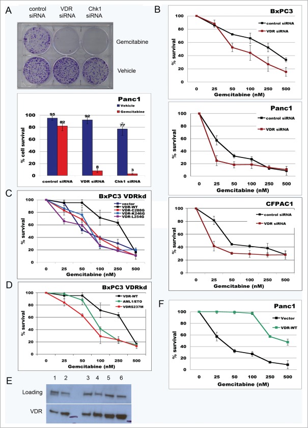
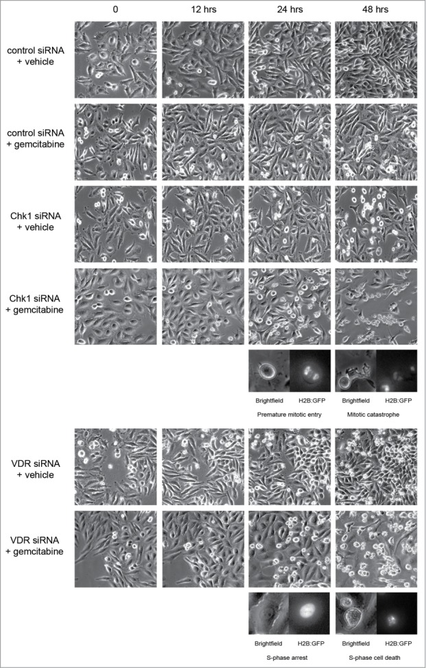
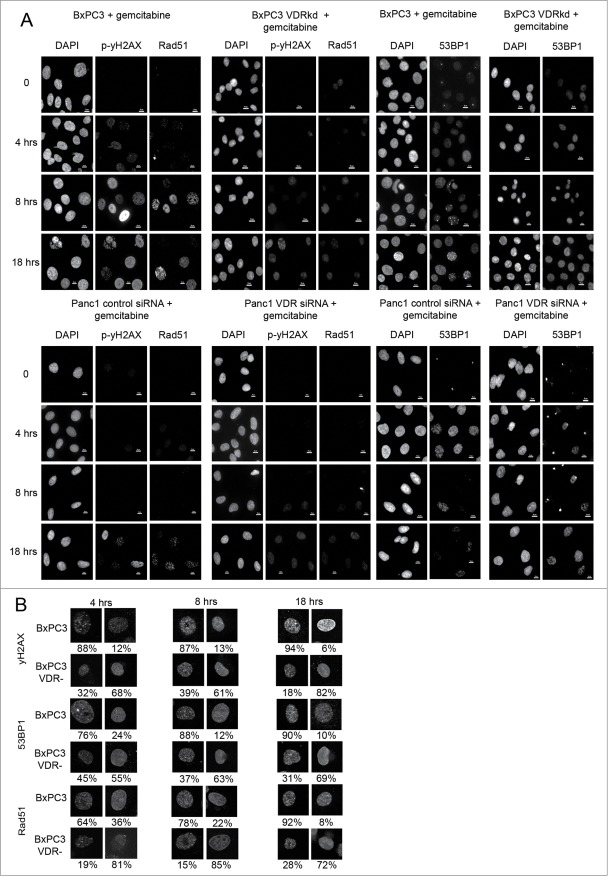
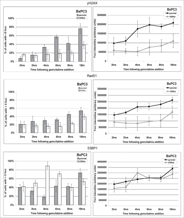
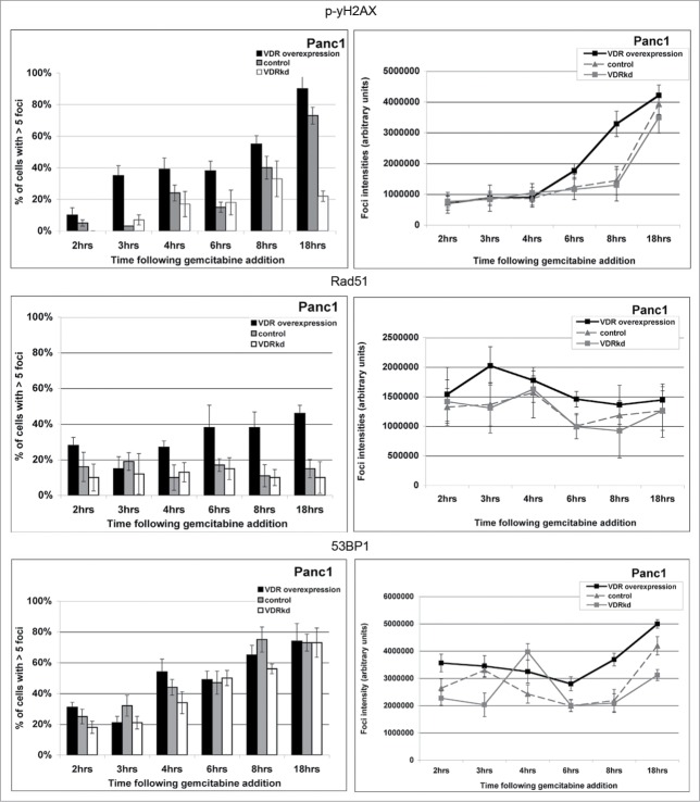
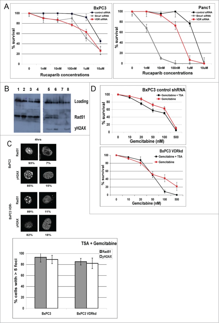
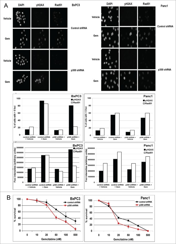
Overcoming chemoresistance of pancreatic cancer (PCa) cells should significantly extend patient survival. The current treatment modalities rely on a variety of DNA damaging agents including gemcitabine, FOLFIRINOX, and Abraxane that activate cell cycle checkpoints, which allows cells to survive these drug treaments. Indeed, these treatment regimens have only extended patient survival by a few months. The complex microenvironment of PCa tumors has been shown to complicate drug delivery thus decreasing the sensitivity of PCa tumors to chemotherapy. In this study, a genome-wide siRNA library was used to conduct a synthetic lethal screen of Panc1 cells that was treated with gemcitabine. A sublethal dose (50 nM) of the drug was used to model situations of limiting drug availability to PCa tumors in vivo. Twenty-seven validated sensitizer genes were identified from the screen including the Vitamin D receptor (VDR). Gemcitabine sensitivity was shown to be VDR dependent in multiple PCa cell lines in clonogenic survival assays. Sensitization was not achieved through checkpoint override but rather through disrupting DNA repair. VDR knockdown disrupted the cells' ability to form phospho-γH2AX and Rad51 foci in response to gemcitabine treatment. Disruption of Rad51 foci formation, which compromises homologous recombination, was consistent with increased sensitivity of PCa cells to the PARP inhibitor Rucaparib. Thus inhibition of VDR in PCa cells provides a new way to enhance the efficacy of genotoxic drugs.

Keywords: BXPC3; DNA DSB, DNA Double-strand break; DNA repair; HDAC inhibitors; IF, Immunofluorescence; PARP inhibitor; PCa, Pancreatic cancer; Panc1; Rad51 foci; VDR; VDR, Vitamin D receptor; Vitamin D receptor; chemosensitization; gemcitabine; p300; pancreatic cancer; siRNA screen; stalled replication fork.


Figures

Figure 1.
Figure 1.
For figure legend, see page 3843.Figure 1 (See previous page). Sensitization of pancreatic cancer cells to gemcitabine following VDR knockdown. (A) Colony formation assay comparing gemcitabine sensitivity of Panc1 cells after control, VDR and Chk1 siRNA transfection. Cells were treated with 50nM gemcitabine for 24 hrs. and drug removed before the asssay. Colony counts are presented beneath the image of a representative colony survival assay. (B) Gemcitabine kill curves from clonogenic survival assays performed on BXPC3, Panc1 and CFPAC1 cells following control or VDR siRNA transfection (n = 5). p values: BxPC3 = 0.036, Panc1 = 0.171, CFPAC1 = 0.083. (C) clonogenic survival assays of gemcitabine sensitivity of BxPC3 VDRkd cells transfected with the indicated VDR constructs (n = 5). p values: VDR-WT = 0.088, VDR-C288G = 0.671, VDR-K246G = 0.845, VDR-L254G = 0.148. (D) AML1/ETO and VDR-S237M neutralizes the ability of WT-VDR to rescue gemcitabine resistance of BxPC3 VDRkd cells (n = 5). p values: AML1/ETO = 0.147, VDR-S237M = 0.039. (E) Western blot showing VDR expression after 18 hour vehicle or gemcitabine (50 nM) treatment of PCa cell lines. 40μg of protein loaded. Lane 1 = Panc1 + Vehicle; Lane 2 = Panc1 + gemcitabine; Lane3 = CFPAC1 + Vehicle; Lane 4 = CFPAC1 + gemcitabine; Lane 5 = BxPC3 + Vehicle; Lane 6 = BxPC3 + gemcitabine. The 55 kDa marker is labeled between lanes 2 and 3, and to the right of lane 6. (F) Increased resistance of Panc1 cells to gemcitabine following VDR overexpression (n = 5). p value = 0.008.
Figure 2.
Figure 2.
Gemcitabine sensitization after VDR knockdown is not due to override of the DNA damage checkpoint. Select frames from a 48 hr timelapse of Panc1 cells transfected with control, Chk1, and VDR siRNAs that were treated with vehicle or gemcitabine. Chk1 siRNA of gemcitabine treated samples show increased mitotic cells at later timepoints. Enlarged images show brightfield and gfpH2B images of Chk1 siRNA cells prematurely entering mitosis and undergoing mitotic catastrophe. VDR siRNA transfected cells remain in interphase and die without ever entering mitosis.
Figure 3.
Figure 3.
For figure legend, see page 3846.Figure 3 (See previous page). VDR knockdwon reduces gemcitabine induced γH2AX and Rad51 foci formation in BXPC3 and Panc1 cells. (A) Cells were treated with 50 nM gemcitabine, fixed at 0, 2 hrs, 3 hrs, 4 hrs, 6 hrs, 8 hrs, and 18 hrs and stained for Rad51, γH2AX, and 53BP1. Representative images (40X) from 0, 4, 8 and 18 hrs post drug treatments are shown. (B) Higher magnification (90x) confocal images of individual nuclei displaying the different staining patterns of Rad51, γH2AX, and 53BP1 after VDR knockdown compared to controls. Percentages of each pattern from 500 cells/sample analyzed are presented.
Figure 4.
Figure 4.
Quantification of Rad51, γH2AX, and 53BP1 staining of BxPC3 cells. Individual nuclei from images in Figure 3 were separately analyzed for foci number and focal intensity. Quantitation was performed on cells treated with gemcitabine for 0, 2 hrs, 3 hrs, 4 hrs, 6 hrs, 8 hrs, and 18 hrs. 500 cells from each timepoint was examined for Rad51, γH2AX, and 53BP1.
Figure 5.
Figure 5.
Quantification of Rad51, γH2AX, and 53BP1 staining of Panc 1 cells. Individual nuclei from images in Figure 3 were separately analyzed for foci number and focal intensity. Quantitation was performed on cells treated with gemcitabine for 0, 2 hrs, 3 hrs, 4 hrs, 6 hrs, 8 hrs, and 18 hrs. 500 cells from each timepoint was examined for Rad51, γH2AX, and 53BP1.
Figure 6.
Figure 6.
For figure legend, see page 3850.Figure 6 (See previous page). VDR knockdown sensitizes BXPC3 and Panc1 cells to the PARP inhibitor, Rucaparib. blocks gemcitabine induced DSB HR repair by reducing Rad51 foci formation at DSBs. (A) Rucaparib kill curves generated from clonogenic assays of cells transfected with control, BRCA1 and VDR siRNAs. (B) Western blot comparing Rad51 and γH2AX protein levels of BxPC3 and BxPC3 VDRkd cells. 40μg of protein loaded. Lane 1 = BxPC3 + vehicle (18 hrs) (supernatant fraction); Lane 2 = BxPC3 + gemcitabine (50 nM) (18 hrs) (supernatant fraction); Lane 3 = BxPC3 VDRkd + vehicle (18 hrs) (supernatant fraction); Lane 4 = BxPC3 VDRkd + gemcitabine (50 nM) (18 hrs) (supernatant fraction); Lane 5 = BxPC3 + vehicle (18 hrs) (pellet fraction); Lane 6 = BxPC3 + gemcitabine (50 nM) (18 hrs) (pellet fraction); Lane 7 = BxPC3 VDRkd + vehicle (18 hrs) (pellet fraction); Lane 8 = BxPC3 VDRkd + gemcitabine (50 nM) (18 hrs) (pellet fraction). (C) Comparison of Rad51 and γH2AX staining of BxPC3 and BxPC3kd cells following TSA (500 nM) + gemcitabine (50 nM) treatments. (D) Colony survival of BxPC3 VDRkd and BxPC3 parental cells treated with TSA (500 nM) + gemcitabine (n = 3). p values: BxPC3 VDRkd = 0.289, BxPC3 control = 0.02.
Figure 7.
Figure 7.
p300 knockdown reduces gemcitabine induced Rad51 foci formation. (A) Rad51 and yH2AX staining of BxPC3 or Panc1 cells treated with control and p300 shRNA's, and treated with vehicle or gemcitabine. (B) Clonogenic survival assay of BxPC3 and Panc1 cells transfected with p300 shRNA and treated with different doses of gemcitabine (n = 3). p values: BxPC3 = 0.0489, Panc1 = 0.192.

References

    1. Caldas C, Kern SE. K-ras mutation and pancreatic adenocarcinoma. Int J Pancreatol 1995; 18:1-6; PMID:7594765 - PubMed
    1. Jones S, Zhang X, Parsons DW, Lin JC, Leary RJ, Angenendt P, Mankoo P, Carter H, Kamiyama H, Jimeno A, et al. Core signaling pathways in human pancreatic cancers revealed by global genomic analyses. Science 2008; 321:1801-6; PMID:18772397; http://dx.doi.org/ 10.1126/science.1164368 - DOI - PMC - PubMed
    1. Ko AH, Tempero MA. Treatment of metastatic pancreatic cancer. J Natl Compr Canc Netw 2005; 3:627-36; PMID:16194454 - PubMed
    1. van Heek T, Rader AE, Offerhaus GJ, McCarthy DM, Goggins M, Hruban RH, Wilentz RE. K-ras, p53, and DPC4 (MAD4) alterations in fine-needle aspirates of the pancreas: a molecular panel correlates with and supplements cytologic diagnosis. Am J Clin Pathol 2002; 117:755-65; PMID:12090425; http://dx.doi.org/ 10.1309/5RQ0-JCQU-5XF2-51LQ - DOI - PubMed
    1. Weinstein IB. Cancer. Addiction to oncogenes–the Achilles heal of cancer. Science 2002; 297:63-4; PMID:12098689; http://dx.doi.org/ 10.1126/science.1073096 - DOI - PubMed

Publication types

MeSH terms