. 2014;13(24):3867-77.
doi: 10.4161/15384101.2014.972882.

Kizuna is a novel mitotic substrate for CDC25B phosphatase

Affiliations

Kizuna is a novel mitotic substrate for CDC25B phosphatase

Yann Thomas et al. Cell Cycle. 2014.

Abstract

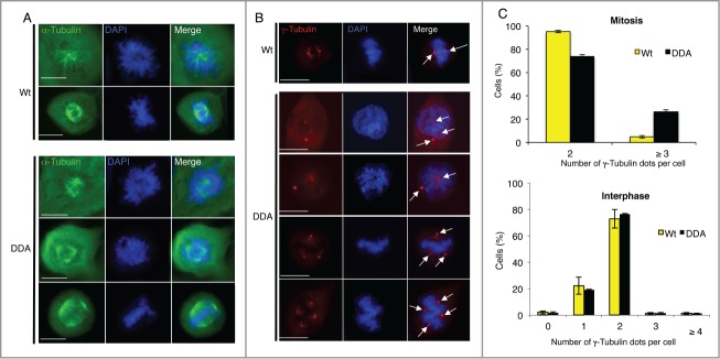
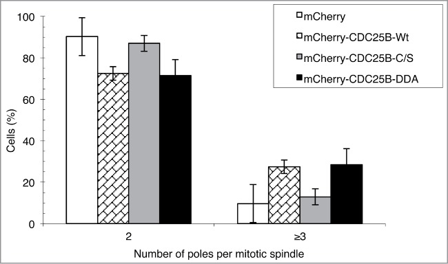
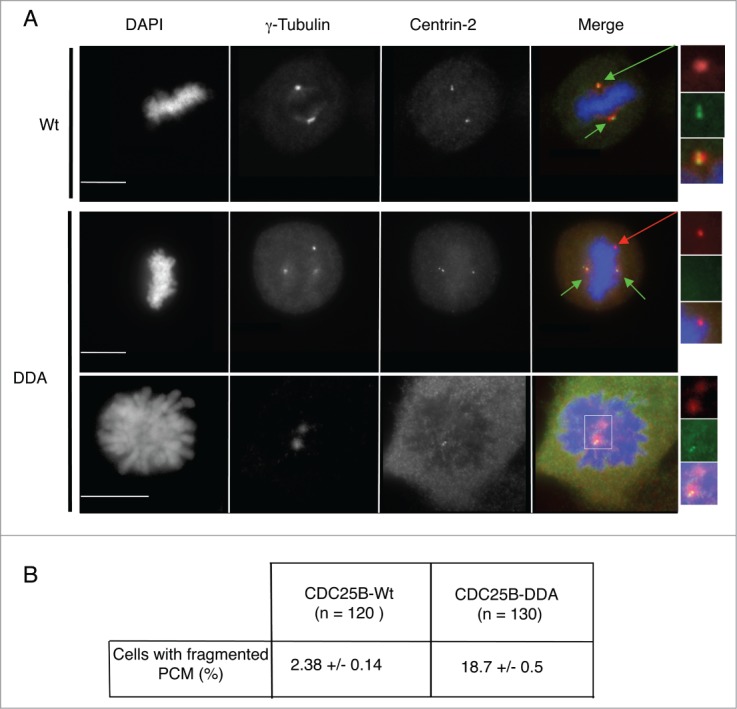
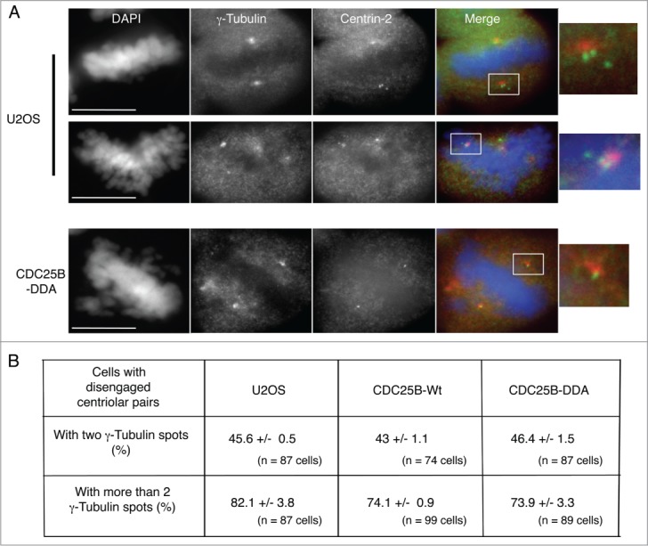
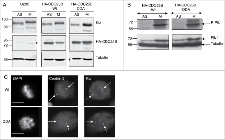
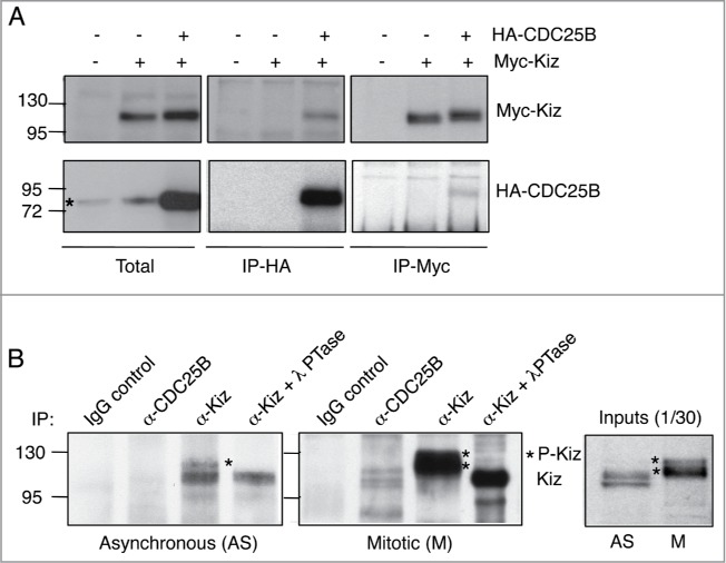
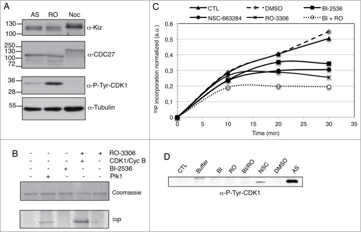
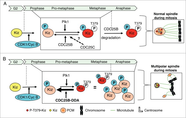
CDC25 dual-specificity phosphatases play a central role in cell cycle control through the activation of Cyclin-Dependent Kinases (CDKs). Expression during mitosis of a stabilized CDC25B mutant (CDC25B-DDA), which cannot interact with the F-box protein βTrCP for proteasome-dependent degradation, causes mitotic defects and chromosome segregation errors in mammalian cells. We found, using the same CDC25B mutant, that stabilization and failure to degrade CDC25B during mitosis lead to the appearance of multipolar spindle cells resulting from a fragmentation of pericentriolar material (PCM) and abolish mitotic Plk1-dependent phosphorylation of Kizuna (Kiz), which is essential for the function of Kiz in maintaining spindle pole integrity. Thus, in mitosis Kiz is a new substrate of CDC25B whose dephosphorylation following CDC25B stabilization leads to the formation of multipolar spindles. Furthermore, endogenous Kiz and CDC25B interact only in mitosis, suggesting that Kiz phosphorylation depends on a balance between CDC25B and Plk1 activities. Our data identify a novel mitotic substrate of CDC25B phosphatase that plays a key role in mitosis control.

Keywords: CDC25B; DMSO, dimethyl-sulfoxyde; DSP, Dithiobis [succinimidyl propionate]; Kiz, Kizuna protein; Kizuna; PCM, pericentriolar material; centrosome; mitosis.


Figures

Figure 1.
Figure 1.
Stabilization of CDC25B causes centrosome aberration only in mitosis. (A). Twenty-4 hours after induction of CDC25B-Wt (Wt) or CDC25B-DDA (DDA) expression in U2OS Tet-off cell lines stably transfected with the respective plasmids, asynchronous cells were fixed, and incubated with an anti-α-Tubulin antibody (green). DNA was stained with DAPI. Images are from fluorescence microscopy of prophasic and metaphasic cells. Scale bar: 10 μm. (B). Experiments were performed as in A, except that cells were stained with an anti-γ-Tubulin antibody (red). Positive γ-Tubulin dots are highlighted by arrows. Images are from fluorescence microscopy of representative prophasic and metaphasic cells (3 independent experiments). Scale bar: 10 μm. (C). Quantification of the number of γ-Tubulin-positive dots per cell in mitotic (upper panel) or interphase (lower panel) U2OS cells that express either CDC25B-Wt or CDC25B-DDA. Error bars indicate ± SD of 3 independent experiments.
Figure 2.
Figure 2.
CDC25B phosphatase activity is required to induce multipolar spindles in cells. HeLa cells were transiently transfected with pmCherry-CDC25B (-Wt, -C/S or -DDA) or pmCherry expressing vectors. Fourteen hours post-transfection, cells expressing mCherry-tagged CDC25B-Wt, CDC25B-C/S, CDC25B-DDA, or mCherry alone were imaged by time-lapse microscopy for 24 hours. The number of poles per mitotic spindle was counted. Data represent the percentage of cells with 2 or more than 2 poles and values are the mean ± SD of 3 independent experiments. Total number of counted cells: mCherry, n = 161; mCherry-CDC25B-Wt, n = 106; mCherry-CDC25B-C/S, n = 126; mCherry-CDC25B-DDA, n = 144.
Figure 3.
Figure 3.
Multipolar spindles in U2OS-CDC25B-DDA cells are the result of PCM fragmentation. (A). Representative fluorescent microscopy images of asynchronously growing U2OS cells in metaphase or prophase that express CDC25B-Wt or CDC25B-DDA fixed and stained with anti-γ-Tubulin and anti-Centrin-2 antibodies. DNA was stained with DAPI. Centrosomes are highlighted by green arrows, and PCM fragmentation by a red arrow (magnified view in insets). In the merged images, yellow shows co-localization of Centrin-2 and γ-Tubulin. Scale bar: 10 μm. (B). The number of cells with fragmented PCM (i.e., cells with an excess of γ-Tubulin dots) was quantified in U2OS-CDC25B-Wt and U2OS-CDC25B-DDA cells. Values are the mean ± SD of 3 independent experiments.
Figure 4.
Figure 4.
Stabilization of CDC25B does not induce a disengagement of centriolar pairs. (A). Representative fluorescent microscopy images of mitotic cells from an asynchronous population of parental or CDC25B-DDA U2OS cells fixed and stained with anti-γ-Tubulin and anti-Centrin-2 antibodies. DNA was stained with DAPI. In the merged images, yellow shows co-localization of Centrin-2 and γ-Tubulin. Scale bar: 10 μm. (B). The number of cells with disengaged centriolar pairs (i.e., cells in which the distance between centriols is greater than expected for a diplosome) in cells presenting either 2 or more than 2 γ-Tubulin dots was quantified in U2OS, U2OS-CDC25B-Wt and U2OS-CDC25B-DDA cells. Values are the mean ± SD of 3 independent experiments.
Figure 5.
Figure 5.
Stabilization of CDC25B during mitosis modifies Kiz phosphorylation without affecting Plk1 activity and centrosomal localization of Kiz. (A). Asynchronously growing (AS) and nocodazole treated (M) parental U2OS cells or that express CDC25B-Wt or CDC25B-DDA were lysed and immunoprobed as indicated. *: non-specific band. (B). Experiments were performed as in A, except that cell extracts were immunoprobed with anti-Plk1 and anti-Phopho-Plk1 (Thr120). (C). Twenty-4 hours after the induction of CDC25B-Wt or CDC25B-DDA expression in U2OS Tet-off cells, cells were fixed and incubated with anti-Centrin-2 and -Kiz antibodies. DNA was stained with DAPI. Images of mitotic cells are shown. Scale bar, 10 μm.
Figure 6.
Figure 6.
Kiz is dephosphorylated by CDC25B, not CDC25A. (A). Recombinant GST-Kiz-Wt or GST-Kiz-TA (T379A) immobilized on Glutathione beads was used in a phosphorylation/dephosphorylation assay in the presence of recombinant His6-Plk1 and MBP-CDC25A or MBP-CDC25B, as described in “Materials and methods." The phosphorylation level of GST-Kiz was then determined by autoradiography. Incorporation of P into GST-Kiz, was normalized to the protein amount of each band based on Coomassie staining (left panel). The relative activity of recombinant MBP-CDC25 phosphatases was indirectly determined by measuring the activation of inactive CDK1 associated with Cyclin B (i.e., CDK1 phosphorylation on Thr14 and Tyr15), in a kinase assay using histone H1 as substrate. For this 50 ng of recombinant MBP-CDC25A or MBP-CDC25-B were incubated with immunoprecipitated inactive CDK1/Cyclin B complexes. Activity of CDK1/Cyclin B complexes was then tested in an in vitro histone H1 kinase assay, as described in. Equal loading was shown by Coomassie staining. The incorporation of P into histone H1 reflects the activity of CDK1/Cyclin B complexes (right panel). (B). Quantification of P incorporation in recombinant GST-Kiz obtained in A. Values from 3 independent experiments were normalized to those of GST-Kiz-Wt incubated only with His6-Plk1. Data are the mean ± SEM of 3 independent experiments (*, P = 0.0318). (C). Quantification ofP incorporation in recombinant GSt-Kiz-Wt during a kinetic of dephosphorylation by recombinant MBP-CDC25B, in presence or not of NSC-663284 (500 nM), or MBP-CDC25A. Values were normalized to those of GST-Kiz-Wt incubated only with His6-Plk1.
Figure 7.
Figure 7.
CDC25B interacts with Kiz during mitosis. (A). HeLa cells were transfected or not with HA-CDC25B-Wt or Myc-Kiz (pRK5) and lysed 24 h after transfection. Cell extracts were incubated with control IgG or anti-Myc/anti-HA antibodies, and immunocomplexes probed for the indicated proteins. *: non-specific band. (B). Asynchronous (left panel) or mitotic (right panel) HeLa cells were lysed in the presence of a cross-linking agent (DSP), immunoprecipitated with anti-CDC25B or anti-Kiz antibodies and immunoprobed with anti-Kiz antibodies. In indicated cases, immunoprecipitates obtained with anti-Kiz antibodies were treated with lambda phosphatase (λPTase, 200 units) 15 min at 30°C before SDS-PAGE (α-Kiz + λPTase).
Figure 8.
Figure 8.
CDK1/Cyclin B, Plk1 and CDC25B regulate Kiz-phosphorylation level during mitosis. (A). Total cell extract (30 μg) of asynchronous (AS), RO-3306 (RO) or nocodazole treated (Noc) HeLa cells were analyzed on SDS-PAGE and immunoprobed as indicated. Anti-CDC27 and anti-P-Tyr-CDK1 were used to check mitosis progression. Anti-Tubulin served as a loading control. (B). Recombinant GST-Kiz-Wt immobilized on Glutathione beads was used in an in vitro kinase assay in the presence of recombinant His6-Plk1 or GST-CDK1/GST-Cyclin B kinases, pre-incubated or not with BI-2536 (250 nM) and RO-3306 (1 μM), respectively. The phosphorylation level of GST-Kiz was then determined by autoradiography. (C). Recombinant GST-Kiz-Wt immobilized on Glutathione beads was used in a phosphorylation assay in the presence of pro-metaphase HeLa cell extract, as described in “Materials and methods," in presence or not of BI-2536 (250 nM), RO-3306 (1 μM), NSC-663284 (1 μM) or DMSO. The phosphorylation level of GST-Kiz was then determined by autoradiography. Incorporation of P into GST-Kiz, was normalized to the protein amount of each band based on Coomassie staining. Values from 3 independent experiments were normalized to those of GST-Kiz-Wt incubated only with pro-metaphase extract. Data are the mean of 3 independent experiments. (D). Pro-metaphase HeLa cell extract was incubated for 30 min at 30°C with kinase buffer in presence of the indicated kinases or phosphatase inhibitors, as in C, analyzed on SDS-PAGE and immunoprobed with anti-P-Tyr-CDK1. Extract from asynchronous HeLa cells was used as control for CDK1 phosphorylation on Tyr-15.
Figure 9.
Figure 9.
Proposed model for the regulation of Kiz phosphorylation during mitosis. (A). During mitosis, phosphorylation level of Kiz results from CDK1/Cyc B kinase activity and a balance between Plk1 kinase and CDC25B phosphatase activities. At the entrance into mitosis Kiz is phosphorylated by CDK1/Cyc B, a priming phosphorylation that facilitates its phosphorylation on Thr379 by Plk1. Until metaphase-anaphase transition, phosphorylation state of Thr379 results from a balance between activities of Plk1 and CDC25B (and maybe CDC25C) to finely control the equilibrium between expansion and stabilization of the PCM around the centrosomes. At the metaphase-anaphase transition, when CDC25B is degraded by the proteasome, Kiz is fully phosphorylated on Thr379 by Plk1. (B). During mitosis, if a defect in proteasome-dependent degradation of CDC25B occurs, such as when the CDC25B-DDA mutant is expressed, the high level of CDC25B maintains Kiz in an hypo-phosphorylated form that hinders its ability to stabilize PCM around the centrosomes during all phases of mitosis, resulting into the formation of multipolar spindles.

Similar articles

Cited by

References

    1. Morgan DO. Principles of CDK regulation. Nature 1995; 374:131-4; PMID:7877684; http://dx.doi.org/10.1038/374131a0 - DOI - PubMed
    1. Boutros R, Lobjois V, Ducommun B. CDC25 phosphatases in cancer cells: key players? Good targets? Nat Rev Cancer 2007; 7:495-507; PMID:17568790; http://dx.doi.org/10.1038/nrc2169 - DOI - PubMed
    1. Boutros R, Dozier C, Ducommun B. The when and wheres of CDC25 phosphatases. Curr Opin Cell Biol 2006; 18:185-91; PMID:16488126; http://dx.doi.org/10.1016/j.ceb.2006.02.003 - DOI - PubMed
    1. Ferguson AM, White LS, Donovan PJ, Piwnica-Worms H. Normal cell cycle and checkpoint responses in mice and cells lacking Cdc25B and Cdc25C protein phosphatases. Mol Cell Biol 2005; 25:2853-60; PMID:15767688; http://dx.doi.org/10.1128/MCB.25.7.2853-2860.2005 - DOI - PMC - PubMed
    1. Lee G, White LS, Hurov KE, Stappenbeck TS, Piwnica-Worms H. Response of small intestinal epithelial cells to acute disruption of cell division through CDC25 deletion. Proc Natl Acad Sci U S A 2009; 106:4701-6; PMID:19273838; http://dx.doi.org/10.1073/pnas.0900751106 - DOI - PMC - PubMed

Publication types